One important
advantage of the present invention is that the method and posterior retractors are used in a minimally invasive orthopedic
hip surgery. A single, small three inch incision is made at the
surgical site on the side on the hip. The method of the present invention, thus, enjoys the benefits of a shorter incision compared to traditional hip
surgery that uses a much longer incision. As one benefit, the patient can recover in a much shorter period of time after a MIS. The recuperation time in the hospital can be a few days and significantly reduce the cost to both the patient and hospital. This shortened time period is extremely important to the patient. Further, MIS is
less invasive and traumatic to the patient. Significantly less
soft tissue is disrupted in a minimally
invasive surgery compared to a traditional hip surgery. Also, the amount of
blood loss is reduced, and patients will require fewer blood transfusions. Further, the length of the scar is significantly smaller, and these
scars are more cosmetically appealing. The incisions themselves heal in a much shorter period of time and are much less painful than a long ten or twelve inch incision. As such, the patient can sooner
return to work or enjoy recreational activities. In short, the patient can more quickly return to a normal way of life.
is that the method and posterior retractors are used in a minimally invasive orthopedic hip surgery. A single, small three inch incision is made at the
surgical site on the side on the hip. The method of the present invention, thus, enjoys the benefits of a shorter incision compared to traditional hip surgery that uses a much longer incision. As one benefit, the patient can recover in a much shorter period of time after a MIS. The recuperation time in the hospital can be a few days and significantly reduce the cost to both the patient and hospital. This shortened time period is extremely important to the patient. Further, MIS is
less invasive and traumatic to the patient. Significantly less soft tissue is disrupted in a minimally
invasive surgery compared to a traditional hip surgery. Also, the amount of
blood loss is reduced, and patients will require fewer blood transfusions. Further, the length of the scar is significantly smaller, and these
scars are more cosmetically appealing. The incisions themselves heal in a much shorter period of time and are much less painful than a long ten or twelve inch incision. As such, the patient can sooner
return to work or enjoy recreational activities. In short, the patient can more quickly return to a normal way of life.
Another important
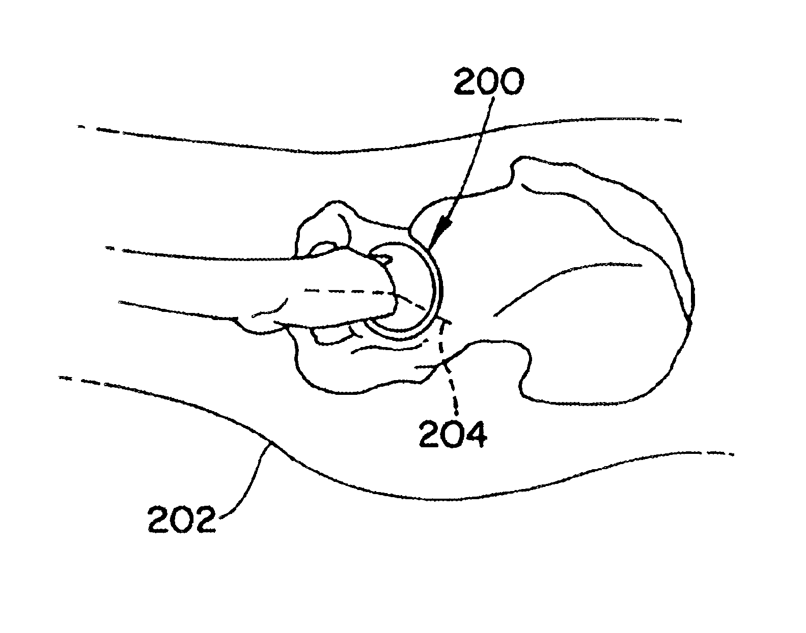
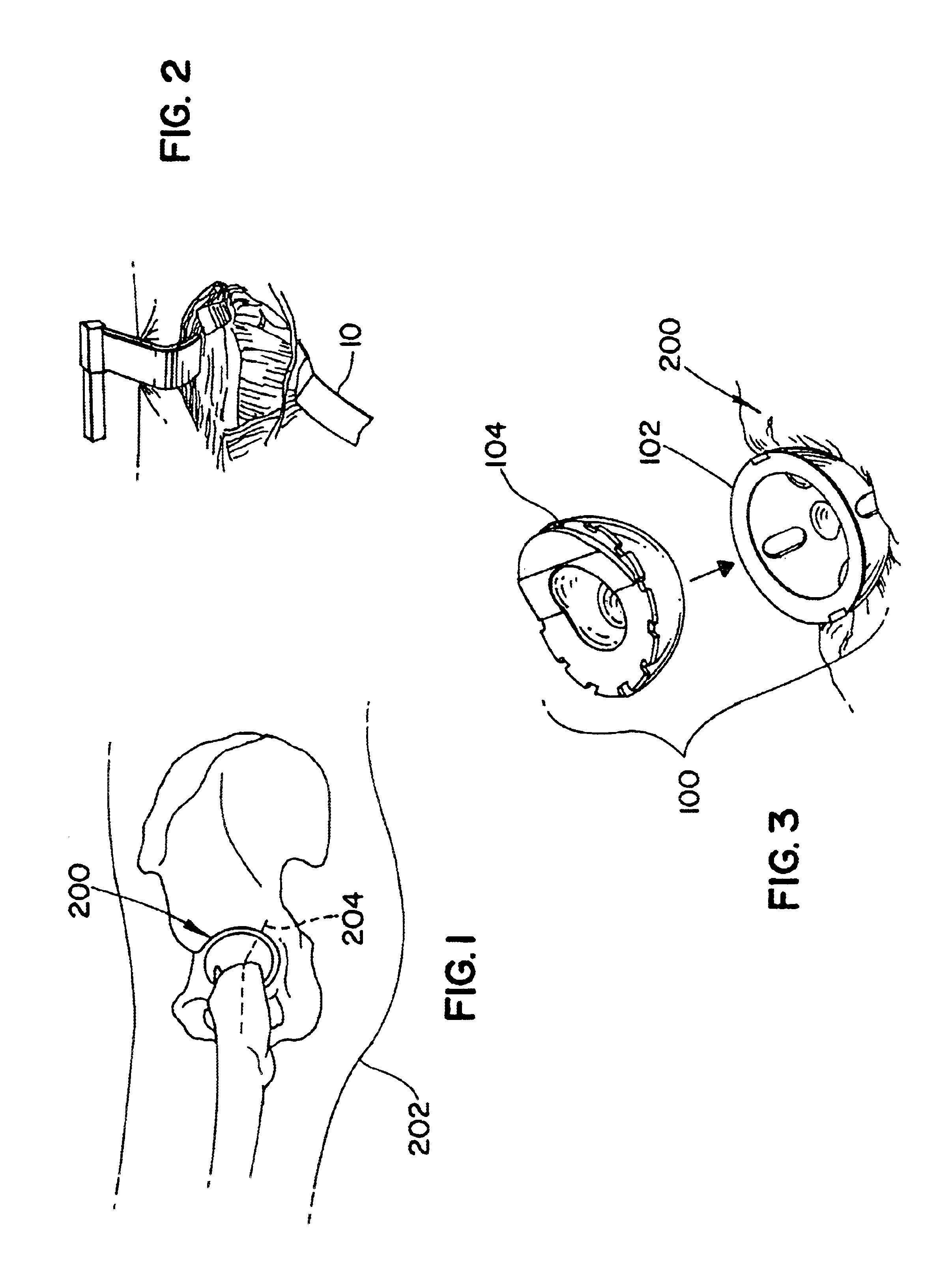
advantage of the present invention is that curved surgical retractors are used in a MIS. The curvature of this instrument and shape of the retracting section are specifically designed and adapted to be used in minimally invasive surgical techniques for retracting soft tissue away from the natural
acetabulum of a patient.
Another important
advantage of the present invention is that the curvature and length of the instrument keep the
handle section away from the entrance to the surgical site. In MIS, it is particularly important to maintain a clear and unobstructed access to the surgical site since it is so small, measuring approximately three to five inches in length. In the present invention, the
handle section of the retractors extends outwardly and away from the surgical site and, thus, does not obstruct access or visual reference to the site.
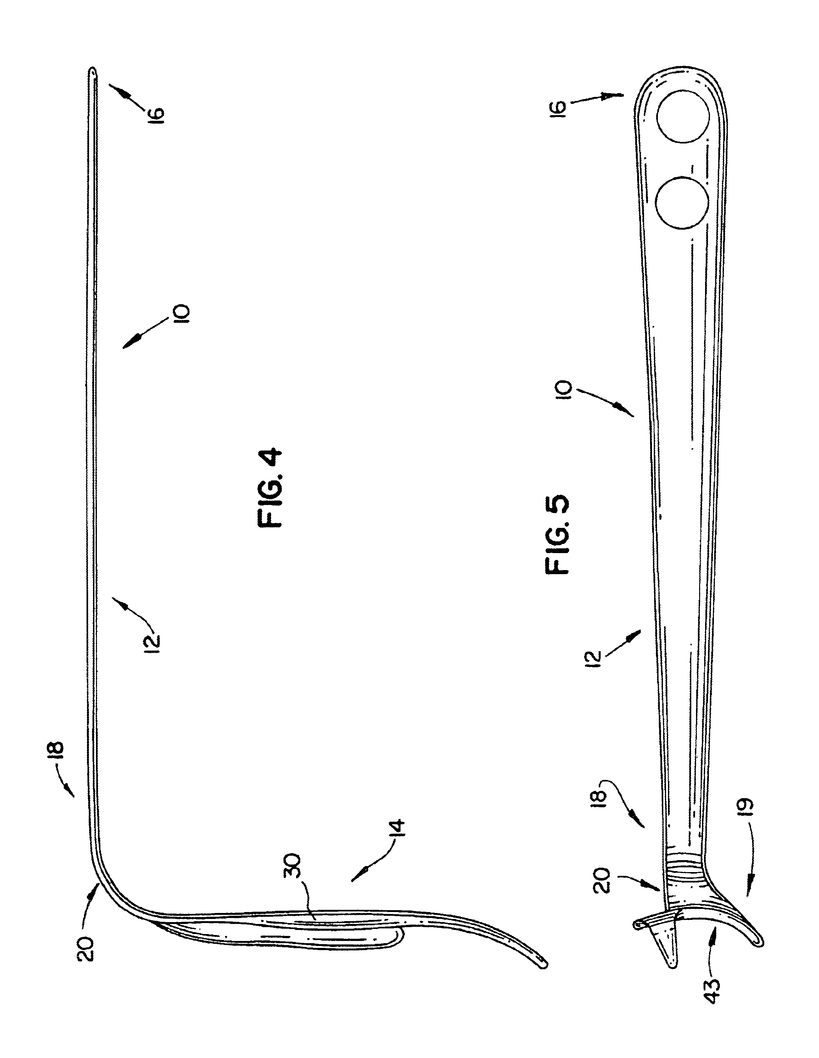
The posterior
retractor generally comprises a body having two primary sections, a handle section and a retracting section. The handle section is elongated with a length specifically adapted to provide sufficient leverage for soft tissue retraction and to position the hand of the user out and away from the surgical site. A distal section of the handle connects to a curved section that leads to the retracting end. This curved section positions the retracting section at about a 90° angle with the handle section. The retracting section includes an elongated
paddle with a straight wall on one side that extends to a curved shoulder and recess located on a distal end. An elongated prong is adjacent the recess and extends upwardly to a
flare that forms a second side opposite to the straight wall. The
flare has a gradual, smooth curvature that transitions inwardly to form the top portion of the
paddle. The paddle includes several critical elements of the invention and is discussed in more detail with reference to the figures.
As another advantage, the posterior retractors can consistently and reliably perform through a small three inch opening in the patient. The shape of the retracting section is specifically sized and shaped to retract and hold tissue in the posterior section of the surgical site. The paddle section, for instance, is designed to rest against the
ischium while keeping soft tissue from sagging backing into the operating area. As another example, the prong is sized and shaped to rest against the obturator fossa so the flared section can push soft tissue out of the surgical site.

