

Method and apparatus of three dimension electrocardiographic imaging
a three-dimensional electrocardiographic and electrocardiographic imaging technology, applied in the field of three-dimensional electrocardiographic imaging, can solve the problems of not providing a single equivalent dipole, and not providing a means of imaging distributed cardiac electrical activity
- Summary
- Abstract
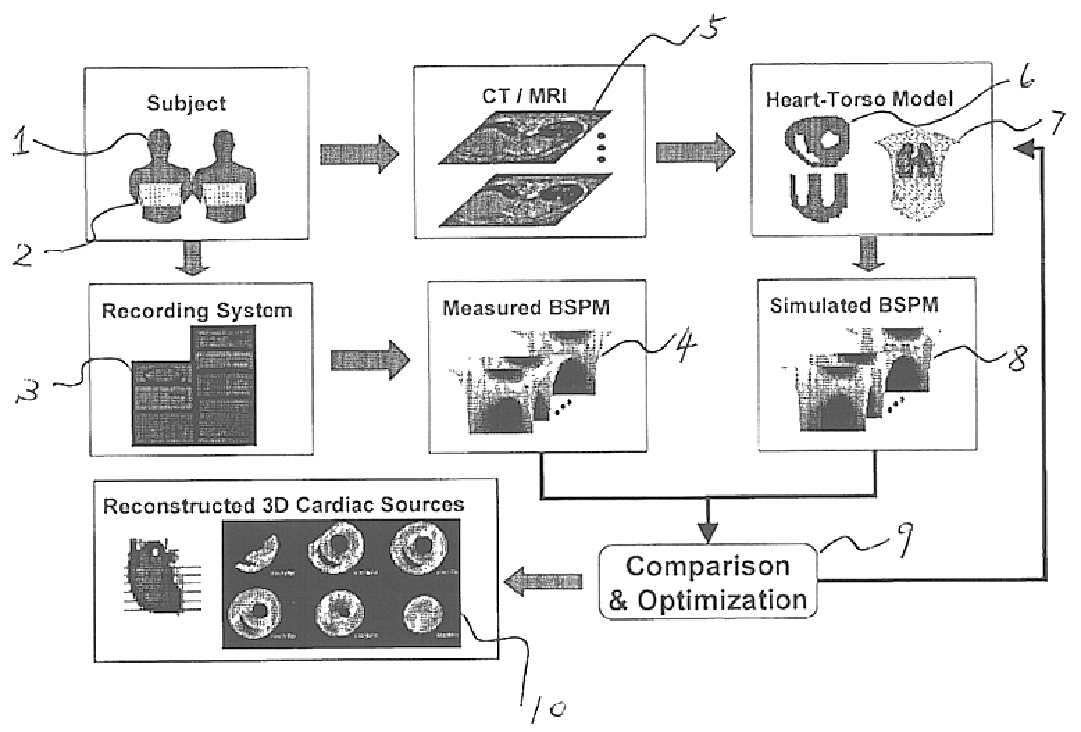
- Description
- Claims
- Application Information
AI Technical Summary
Benefits of technology
Problems solved by technology
Method used
Image

Examples
example i
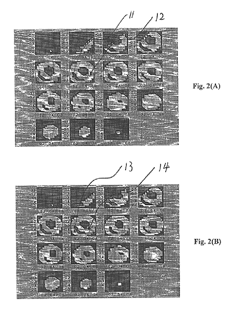
FIG. 2A and FIG. 2B illustrate an example of applying the present invention in localizing and imaging focal sources in the myocardium from body surface electrocardiographic potentials. A three-dimensional heart-torso inhomogeneous volume conductor model is used in this example. The ventricles are divided into an equi-distant lattice structure with an inter-lattice distance of 6.7 mm. In total there are 1,124 voxel nodes in the three dimension solution space of the ventricles. A current dipole located at the right ventricle 11 and a current dipole located at the left ventricle 12, both oriented from the waist towards the neck, are used to approximate well-localized myocardial electrical sources, as shown in FIG. 2A. Gaussian white noise of 5% is added to the calculated body surface potentials from assumed cardiac sources to simulate noise-contaminated body surface electrocardiographic potential measurements. The cardiac source distribution is approximated by a current dipole distribu...
example ii
FIGS. 3-5 illustrate an example of applying the present invention in localizing and imaging site of origin of activation in the myocardium from body surface electrocardiographic potentials in a patient. FIG. 3 shows the boundary element torso model 15 and a finite element ventricle model 16 constructed from contrast and non-contrast ultrafast cardiac computer tomography scans of a male patient with a pacemaker. The realistic-geometry ventricle model 16 consists of 11,144 cubic myocardial cell units with grid resolution of 3 mm. The pacemaker lead tip is identified from the computer tomography images at the lower-anterior region of the right ventricle free wall and close to the septum. The BSPMs between 20 ms and 40 ms following pacing are recorded over 96 sites over the body surface, and used to reconstruct the activation sequence within the ventricles. FIG. 4 illustrates the estimated intracardiac activation patterns 17 at three different layers within the ventricle model 16, which...
PUM
 Abstract
Description
Claims
Application Information