Classifying cells based on information contained in cell images

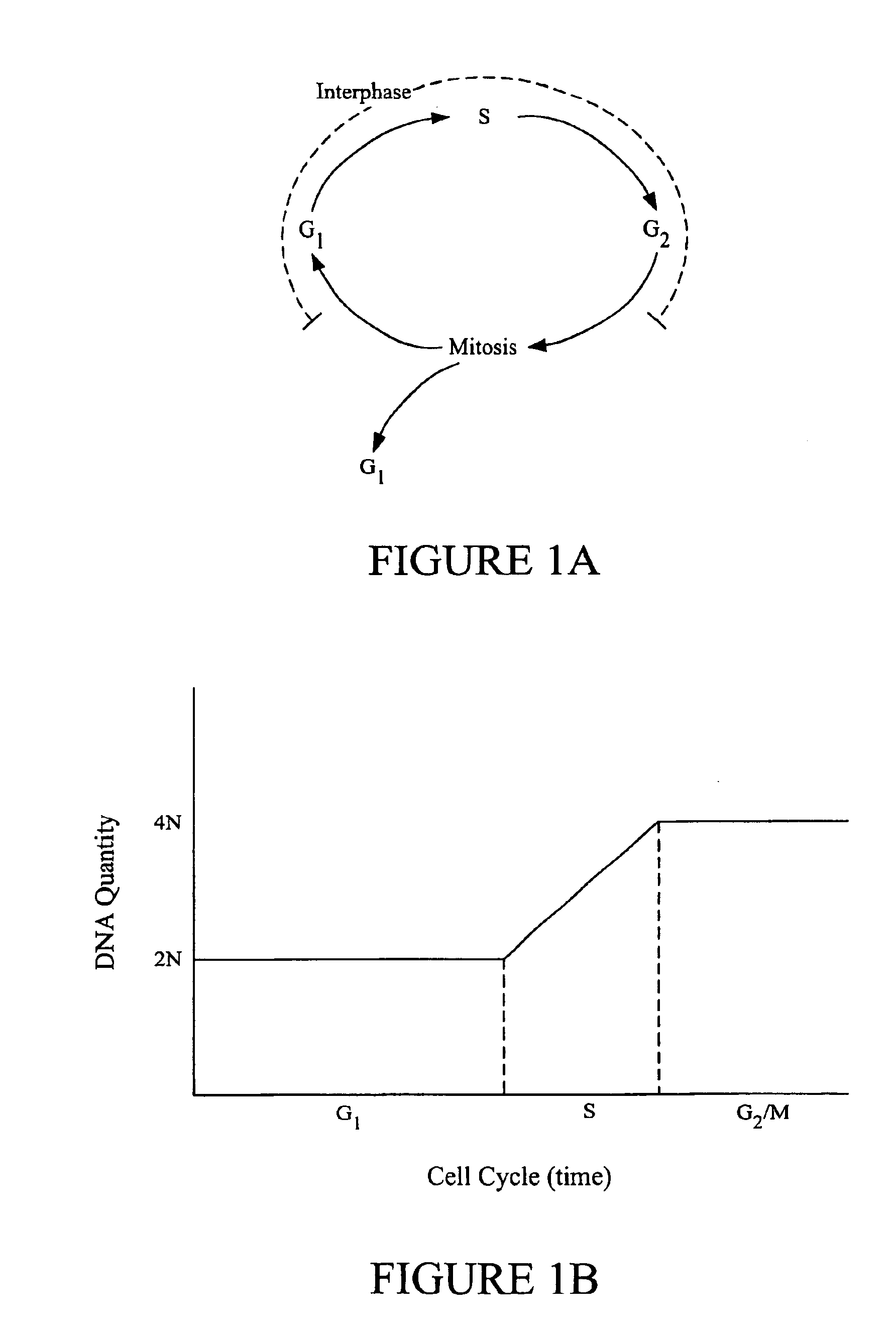
a cell image and information technology, applied in the field of image analysis methods, can solve the problems of inability to distinguish between mitotic cells and gsub>2/sub>phase cells, information provided by fluorescence analyzers is too coarse for many applications,

Inactive Publication Date: 2005-04-05
CYTOKINETICS INC
View PDF34 Cites 66 Cited by
  • Summary
  • Abstract
  • Description
  • Claims
  • Application Information

AI Technical Summary

Benefits of technology

The present invention addresses this need by providing methods and apparatus that analyze images of cells and categorize the cells in particular cell cycle phases based upon certain features extracted from the images. Independently of this, the invention can quantify the total amount of DNA in a cell based on specific features of an image such the as fluorescence intensity generated from fluorescent molecules that bind to DNA. The invention can also characterize a cell as mitotic or interphase based on morphological and textual parameters such as the variance of the pixel intensities observed in a cell image and / or the size of a region containing DNA. Of particular interest, the image analyss methods of this invention can accurately classify a cell into one of the following five phases: G1, S, G2, early mitotic stages (prophase, prometaphase, metaphse), and late mitotic stages (anaphase and telophase).

Problems solved by technology

Unfortunately, information provided by the fluorescence analyzer is too coarse for many applications.
Most importantly, the analyzer only reads out total DNA content per cell; it is unable to distinguish between mitotic cells and G2 phase cells.

Method used


Image

  • Classifying cells based on information contained in cell images
  • Classifying cells based on information contained in cell images
  • Classifying cells based on information contained in cell images

Examples


examples

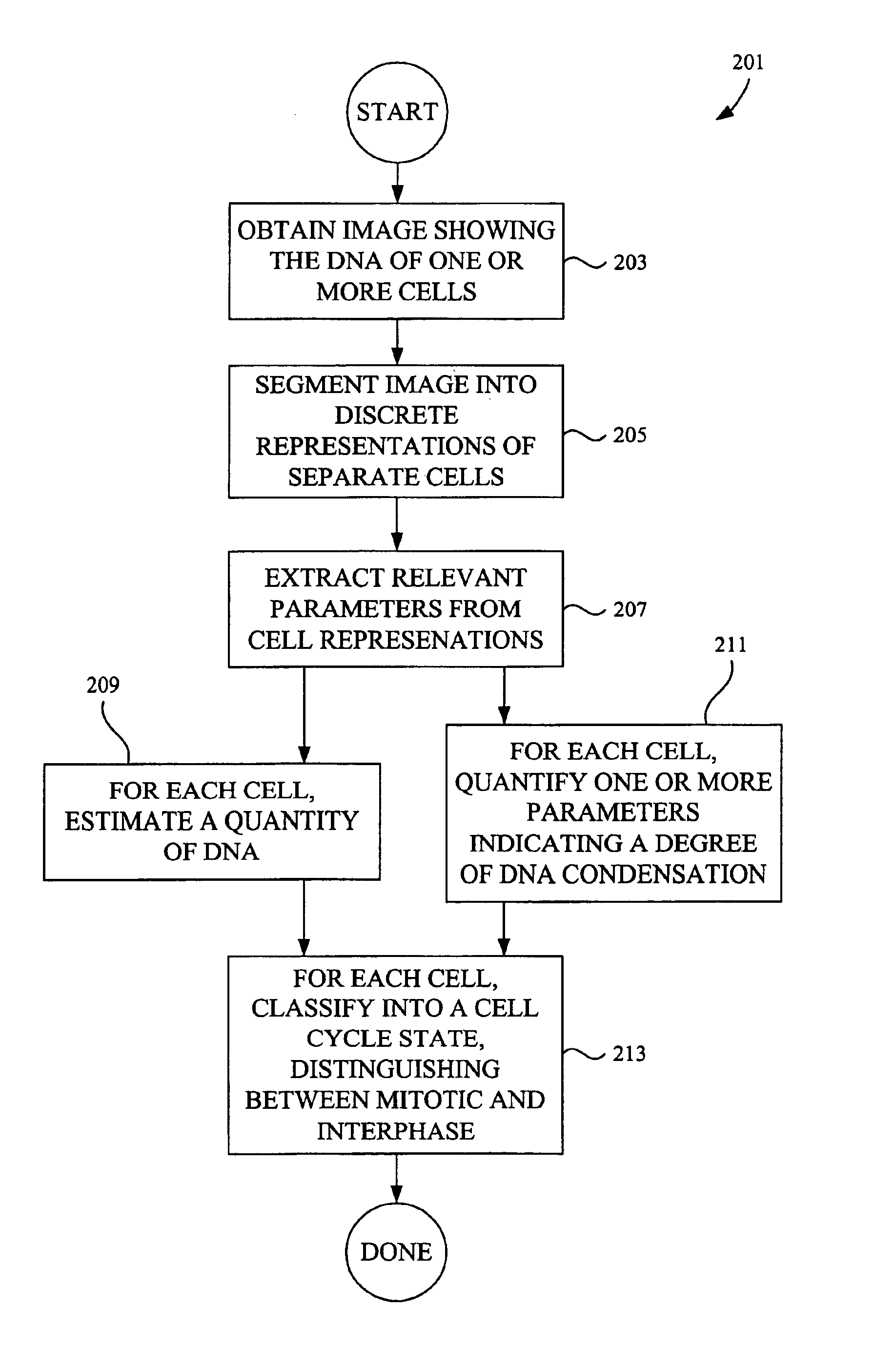
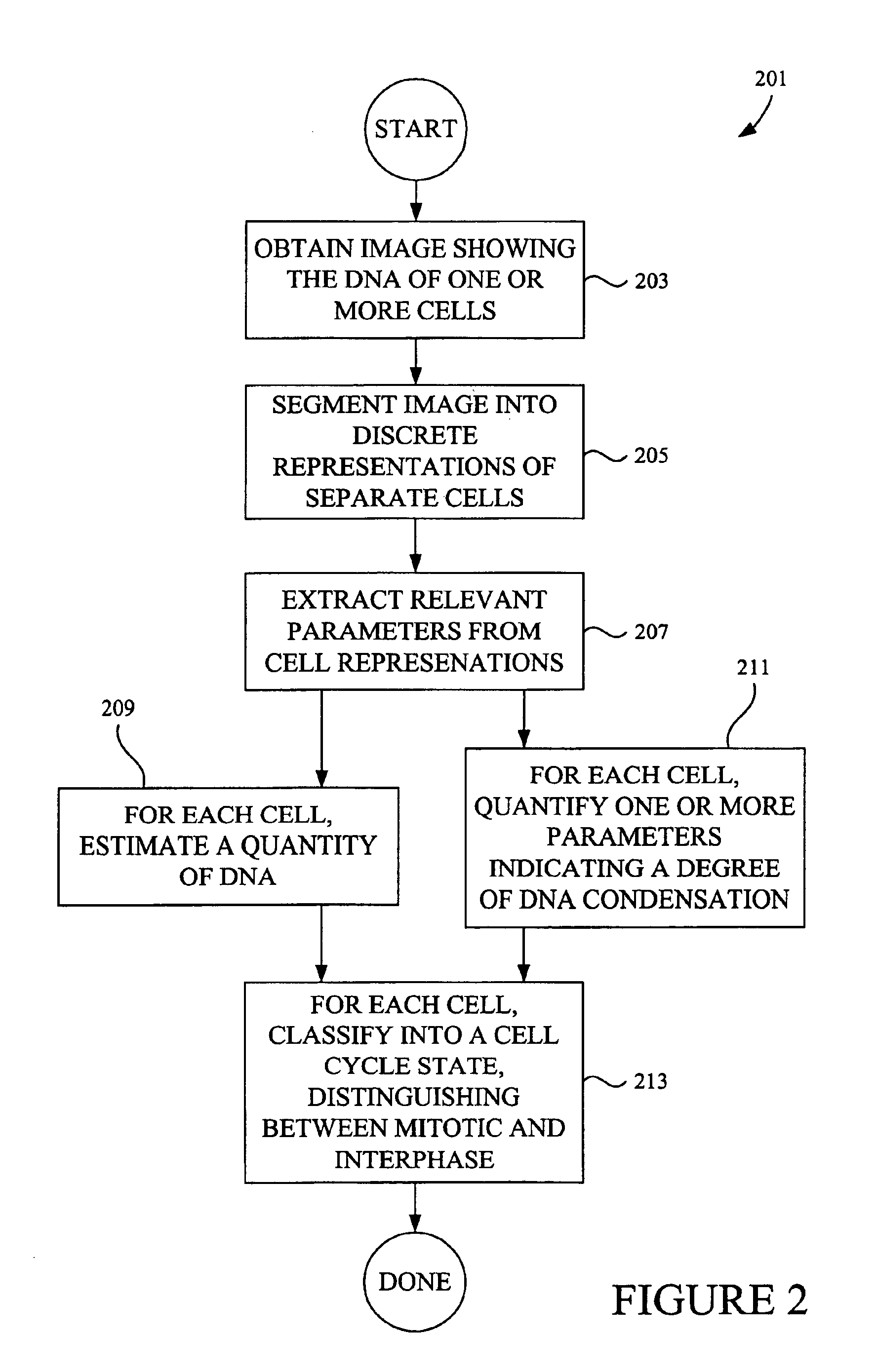
FIGS. 14A-14G depict the results of an experiment in which human lung cancer epithelial cells were treated with Taxol and the effect of Taxol on the cell cycle was characterized. The specific cells used in this study were from Cell Line A549 (human lung cancer epithelial cells) (ATCC:CCL-185). Two day staged cell cultures of the A549 cells were trypsinized for five minutes from T175 cm flasks. The cells were then suspended in ten milliliters of RPMI media with ten percent serum and counted using both a hemocytometer and Coulter Counter. The suspension was diluted further in media to ensure that there were 1600 cells per twenty microliters in suspension. The cells were then counted again. Thereafter, they were transferred to a Cell Stir and kept in suspension while being plated into barcoded 384 well plates using a MultiDrop at twenty microliters per well. The plates were transferred to a humidified carbon dioxide incubator to recover for twenty-four hours before the addition of Taxo...


PUM

PropertyMeasurementUnit
wavelengthaaaaaaaaaa
range of wavelengthsaaaaaaaaaa
interfaceaaaaaaaaaa

Abstract

Image analysis methods analyze images of cells and place the cells in particular cell cycle phases based upon certain features extracted from the images. The methods can also quantify the total amount of DNA in a cell based on specific features such as fluorescence intensity from fluorescent molecules that bind to DNA. Further, the methods can characterize a cell as mitotic or interphase based on chosen parameters such as the variance in intensity observed in a cell image and / or the size of a region containing DNA. In one example, image analysis methods can classify the cell into one of the following five phases: G1, S, G2, telophase, and an early stage mitotic phase comprised of prophase, metaphase, and anaphase.

Description

BACKGROUND OF THE INVENTIONThe present invention pertains to image analysis methods used to classify cells based upon their state in the cell grow th and division cycle.Many interesting bio logical conditions affect or are affected by changes in cell cycle status. For example, some particular condition may cause cells to divide less frequently than normal. Another condition may cause cells to reside in the DNA synthesis state for longer than a normal amount of time. Biological “conditions” of interest to researchers include disease states, normal unperturbed states, quiescent states, states induced by exogenous agents, etc. Valuable insight may be gained by inducing a biological condition through a genetic manipulation, exposure to a particular agent (e.g., a compound, radiation, a field, etc.), deprivation of required substance, and other perturbations.In drug discovery work, valuable information can be obtained by understanding how a potential therapeutic affects cell growth and d...

Claims


Application Information

Patent Timeline
Patent Type & Authority Patents(United States)
IPC IPC(8): G06T5/00G06K9/00G06T7/60
CPCG06K9/0014G06T7/0081G06T7/60G06T2207/30024G06T2207/10056G06T2207/10064G06T7/11G06V20/695
Inventor VAISBERG, EUGENI A.COLEMAN, DANIEL A.
Owner CYTOKINETICS INC
Why Eureka
Try Eureka