
Subdural evacuating port system

Inactive Publication Date: 2005-08-02
MEDTRONIC PS MEDICAL
View PDF42 Cites 22 Cited by
  • Summary
  • Abstract
  • Description
  • Claims
  • Application Information

AI Technical Summary

Benefits of technology

[0012]In view of the foregoing disadvantages inherent in the known techniques and systems for removing fluids from the subdural region of a patient now present in the prior art, the present invention provides a new subdural evacuating port system with a device and method of use wherein the same can be utilized for removing subdural fluid accumulations in a manner that is minimally invasive and promotes the decompression, expansion, and recovery of the brain.
[0014]The invention is especially effective when used on patients having a subdural space filled with fluid that is substantially liquid without significant coagulation of the fluid, including acute patients that are taking anticoagulants to enhance the fluidity of the matter that has accumulated in the subdural space.
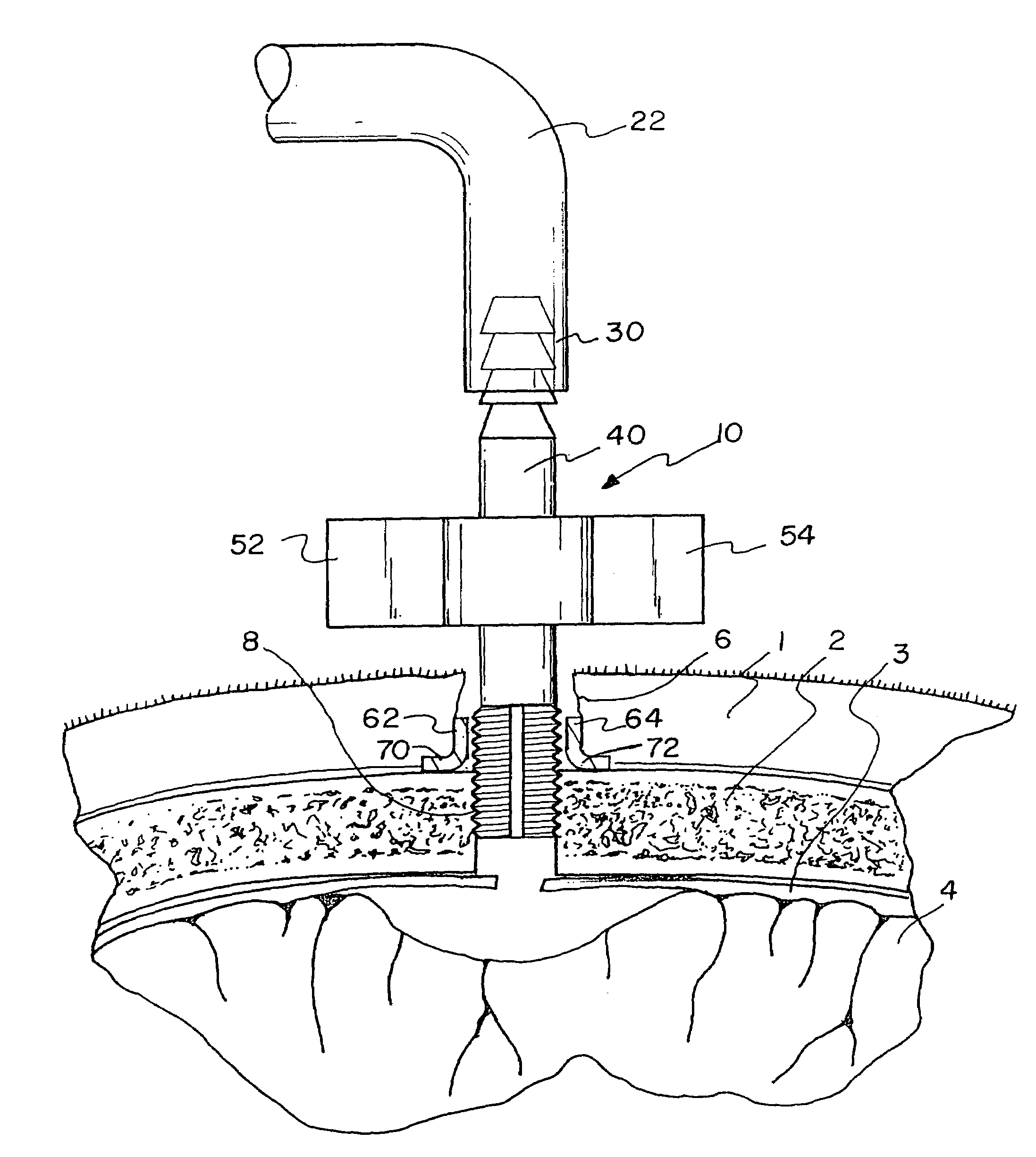
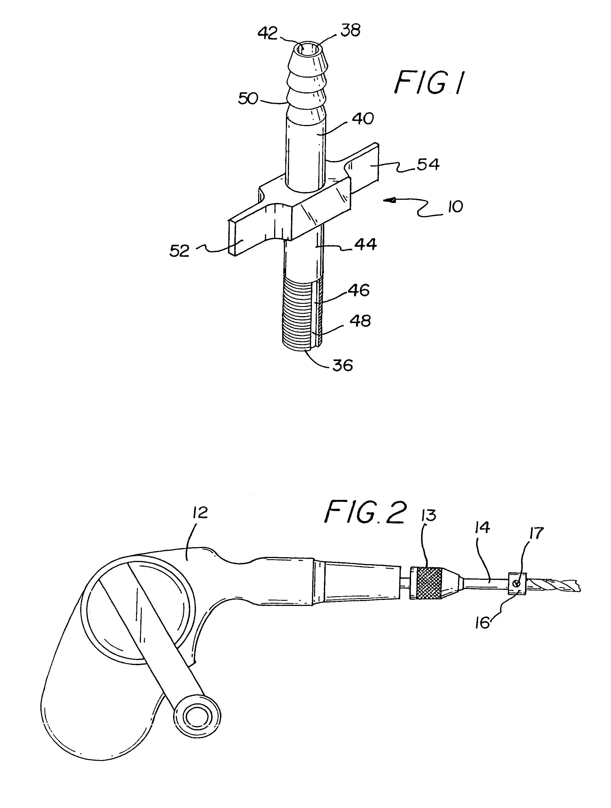
[0015]The present invention generally comprises a subdural evacuating port device including a tubular portion for partial insertion into an opening in a skull of a patient. The tubular portion has a proximal end and a distal end and a lumen extending between the proximal and distal ends. The port device has a pair of wings for facilitating finger rotation of the tubular portion, with the wings extending outwardly from the tubular portion in substantially opposite directions from the tubular portion. The exterior surface of the tubular portion may have self-tapping threads at the proximal end and may have a plurality of annular barbs at the distal end. A kit for evacuating a collection of fluid from a subdural space may include the subdural evacuating port device and a retractor for spacing sides of an incision in a scalp away from each other. The kit may include a drill bit and a stop collar selectively lockable in a position on the drill bit. A method of evacuating a fluid collection from a subdural space includes penetrating the skull to form an opening in the skull, providing the subdural evacuating port device, introducing the proximal end of the subdural evacuating port device into the opening, and creating a substantially uniform negative pressure condition in the subdural space of the patient through the subdural evacuating port device.

Problems solved by technology

For example, elderly persons may be more susceptible to subdural hemorrhages because as the brain ages it tends to become atrophic and the subdural space between the brain and the dura gradually enlarges.
Further, severe linear deceleration of the brain can result in the brain moving excessively with respect to the dura, often causing rupture of the bridging veins or the blood vessels on the surface of the brain, which can in turn cause subdural hemorrhages in the “normal”, young, and otherwise healthy brain.
The craniotomy is a highly invasive procedure that generally involves significant risk to the patient and an extended recovery period.
Again, the patient is exposed to a fairly invasive procedure and a relatively long recovery period.
The typical level of the negative pressure applied by the drains frequently causes further hemorrhage of the brain, especially if the end of the tube should come in contact with the surface of the brain.
Further, recurrence of subdural hematomas and hygromas is quite common in chronic cases as the brain generally fails to expand to fill the enlarged subdural space created by the collection of fluid.
The aforementioned treatment techniques do not actively contribute to re-expansion of the brain within the dura, and therefore do little to prevent the re-accumulation of fluid in the enlarged subdural space.

Method used


Image

  • Subdural evacuating port system
  • Subdural evacuating port system
  • Subdural evacuating port system

Examples


Embodiment Construction

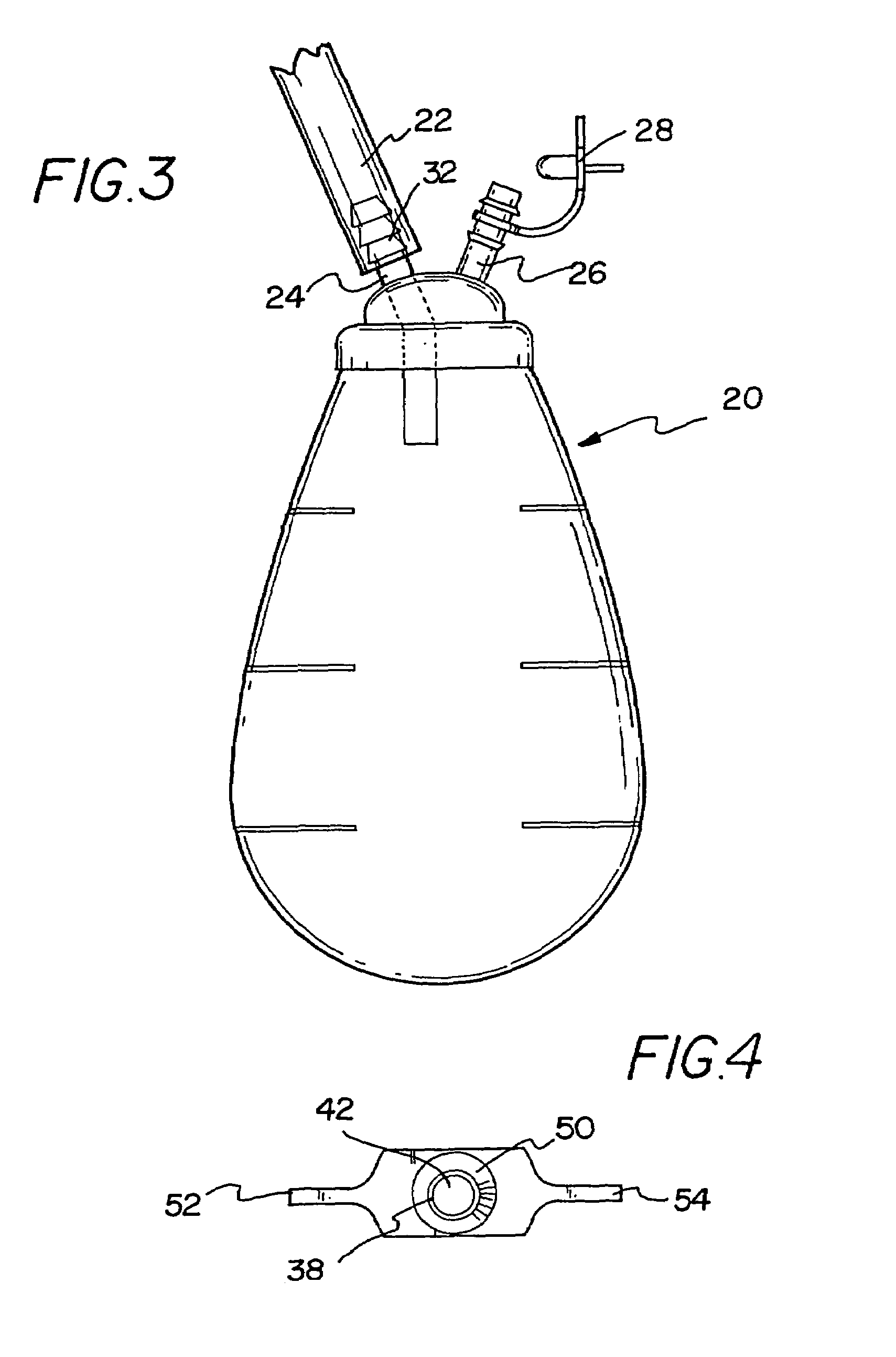
[0028]With reference now to the drawings, and in particular to FIGS. 1 through 7 thereof, a new subdural evacuating port system embodying the principles and concepts of the present invention will be described.

[0029]As best illustrated in FIGS. 1 through 7, the system of the invention generally includes a subdural evacuating port device 10, and contemplates a kit for evacuating a collection of fluid from a subdural space of a patient that incorporates the subdural evacuating port device. The system also contemplates a method for utilizing the subdural evacuating port device and elements of the kit for removing fluid from the subdural space while facilitating the recovery of the patient's brain.

[0030]Elements useful in practicing the invention include the subdural evacuating port device 10, a drill device 12, a drill bit 14 for mounting on the drill device, a stop collar 16 for mounting on the drill bit, a retractor device 18, a negative pressure source 20, and a conduit 22.

[0031]The ...


PUM


Abstract

A subdural evacuating port system for removing subdural fluid accumulations in a manner that is minimally invasive and promotes decompression, expansion and recovery of the brain. The system includes a subdural evacuating port device including a tubular portion for partial insertion into an opening in a skull of a patient. The tubular portion has a proximal end and a distal end and a lumen extending between the proximal and distal ends. The port device has a pair of wings for facilitating finger rotation of the tubular portion, with the wings extending outwardly from the tubular portion in substantially opposite directions from the tubular portion. The exterior surface of the tubular portion may have self-tapping threads at the proximal end and may have a plurality of annular barbs at the distal end. A kit for evacuating a collection of fluid from a subdural space may include the subdural evacuating port device and a retractor for spacing sides of an incision in a scalp away from each other. The kit may include a drill bit and a stop collar selectively lockable in a position on the drill bit. A method of evacuating a fluid collection from a subdural space includes penetrating the skull to form an opening in the skull, providing the subdural evacuating port device, introducing the proximal end of the subdural evacuating port device into the opening, and creating a substantially uniform negative pressure condition in the subdural space of the patient through the subdural evacuating port device.

Description

CROSS REFERENCE TO RELATED APPLICATIONS[0001]This application is a divisional of application Ser. No. 09 / 633,573, Aug. 4, 2000 which is a continuation-in-part of Application No. 29 / 105,951, filed Jun. 4, 1999 now U.S. Pat. No. D435,291.BACKGROUND OF THE INVENTION[0002]1. Field of the Invention[0003]The present invention relates to systems for removing fluids from the subdural region of a patient and more particularly pertains to a new subdural evacuating port system for removing subdural fluid accumulations in a manner that is minimally invasive and promotes decompression, expansion, and recovery of the brain.[0004]2. Description of the Prior Art[0005]The subdural space of the human head is the space located between the brain and the lining of the brain, which is referred to as the dura mater (hereinafter referred to as the “dura”). Hemorrhages on the surface of the brain, for example, may cause a condition known as a subdural hematoma. The subdural hemorrhages may have a number of ...

Claims


Application Information

Patent Timeline
IPC IPC(8): A61M27/00A61M1/00A61B17/16A61B17/02
CPCA61M1/0003A61M1/0023A61M27/006A61M39/0247A61M1/0072A61B17/0218A61B17/1695A61M2039/025A61M2039/0276A61M2039/0291A61M1/82A61M1/60A61M1/98
Inventor ASFORA, WILSON T.
Owner MEDTRONIC PS MEDICAL
Learn More