Methods, apparatus, and computer readable mediums for performing perfusion studies

a perfusion study and computer-readable medium technology, applied in tomography, instruments, applications, etc., can solve the problems of difficult to remove the whole image, high noise, and the sequence of noisy images

Inactive Publication Date: 2005-08-23
UNIV OF IOWA RES FOUND +1
View PDF9 Cites 11 Cited by
  • Summary
  • Abstract
  • Description
  • Claims
  • Application Information

AI Technical Summary

Problems solved by technology

Because a low milliampereage results in higher noise in the projection data, one will get a sequence of noisy images.
However, once the noise is introduced into the collected projection data, it is difficult to remove it completely.

Method used


Image

  • Methods, apparatus, and computer readable mediums for performing perfusion studies
  • Methods, apparatus, and computer readable mediums for performing perfusion studies
  • Methods, apparatus, and computer readable mediums for performing perfusion studies

Examples


Embodiment Construction

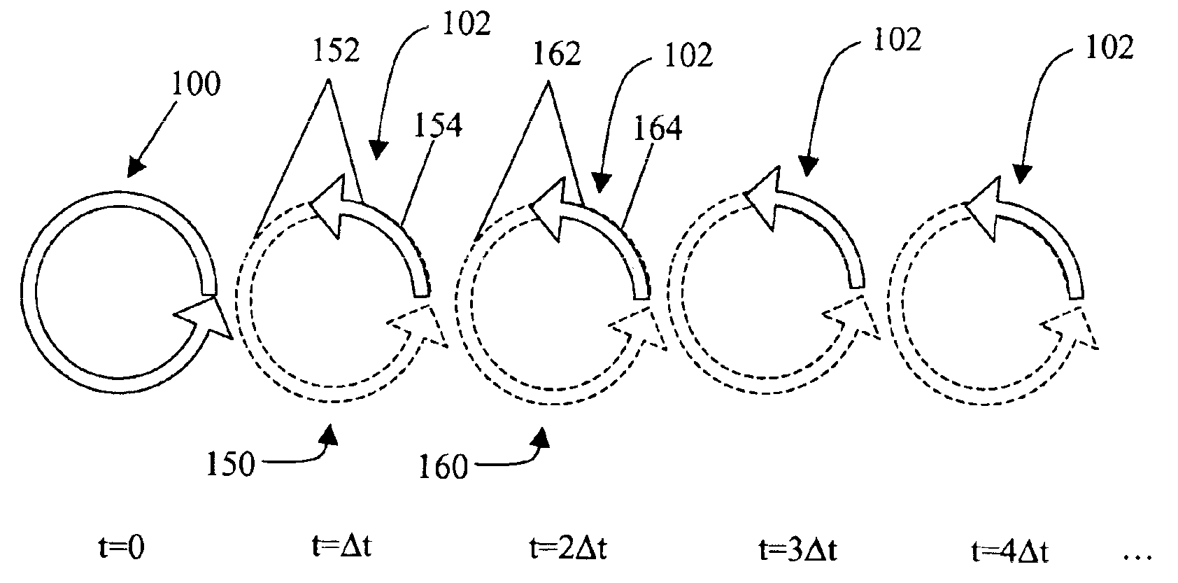
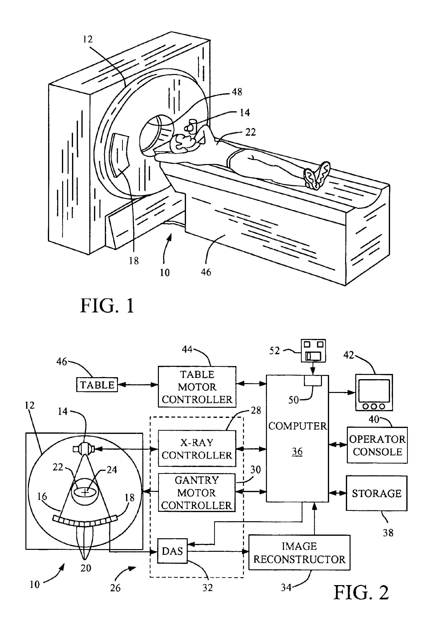
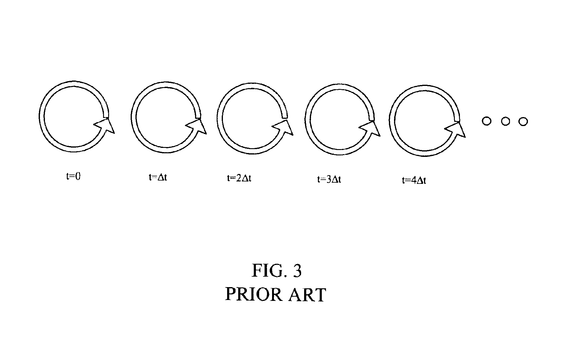
[0020]In some known CT imaging system configurations, an x-ray source projects a fan-shaped beam which is collimated to lie within an X-Y plane of a Cartesian coordinate system and generally referred to as an “imaging plane”. The x-ray beam passes through an object being imaged, such as a patient. The beam, after being attenuated by the object, impinges upon an array of radiation detectors. The intensity of the attenuated radiation beam received at the detector array is dependent upon the attenuation of an x-ray beam by the object. Each detector element of the array produces a separate electrical signal that is a measurement of the beam intensity at the detector location. The intensity measurements from all the detectors are acquired separately to produce a transmission profile.

[0021]In third generation CT systems, the x-ray source and the detector array are rotated with a gantry within the imaging plane and around the object to be imaged such that the angle at which the x-ray beam ...


PUM

PropertyMeasurementUnit
fan angleaaaaaaaaaa
fan angleaaaaaaaaaa
timeaaaaaaaaaa

Abstract

A method for conducting a perfusion study includes performing an initial full scan of an area of interest in an object, and performing at least one subsequent partial scan of the area of interest to detect motion of a contrast agent.

Description

BACKGROUND OF THE INVENTION[0001]This invention relates generally to Computed Tomography (CT) imaging systems, and more particularly to projection interpolation algorithms and image prediction algorithms for low dose perfusion technology.[0002]CT perfusion is a recent advancement in clinical applications. In such a scan, the organ-of-interest is repeatedly scanned in a cine mode (table remains stationary during the scan) while the contrast medium is injected into the patient and propagated in the blood circulation. By monitoring the contrast uptake of the different part of the organ, the mean transit time (MTT), cerebral blood flow (CBF), cerebral blood volume (CBV), and other parameters can be calculated. These parameters are used to differentiate viable versus non-viable tissues. In the current protocol, the patient is continuously scanned at one-second intervals for 30 to 40 second(s).[0003]It is desirable to reduce the amount of dose to the patient. One known way is to reduce th...

Claims


Application Information

Patent Timeline
Patent Type & Authority Patents(United States)
IPC IPC(8): A61B6/03A61B6/00G01N23/02G01N23/04
CPCA61B6/032A61B6/481A61B6/507G01N23/046A61B6/027Y10S378/901G01N2223/419
Inventor WANG, GEHSIEH, JIANGWEI, YUCHUAN
Owner UNIV OF IOWA RES FOUND
Why Eureka
Try Eureka