Offset proximal cage for embolic filtering devices

a filter device and proximal cage technology, applied in the field of filtering devices, can solve the problem of virtually unobstructed filter opening, achieve good flexibility and bendability, enhance the flexibility and bendability of the embolic filter device, and enhance the wall apposition of the filter

Inactive Publication Date: 2005-09-06
ABBOTT CARDIOVASCULAR
View PDF10 Cites 160 Cited by
  • Summary
  • Abstract
  • Description
  • Claims
  • Application Information

AI Technical Summary

Benefits of technology

[0014]The present invention provides a flexible cage or basket for use with an embolic filtering device designed to capture embolic debris created during the performance of a therapeutic interventional procedure, such as a balloon angioplasty or stenting procedure, in a body vessel. The present invention provides the physician with an embolic filtering device having good flexibility to allow it to be steered through tortuous anatomy, but yet possesses sufficient strength to hold open a filtering element against the wall of the body vessel for capturing embolic debris. The present invention creates an embolic filtering device that can be fully deployed within a body vessel and can provide a virtually unobstructed opening for the filtering element which captures embolic particles entrained in the body fluid. An embolic filtering device made in accordance with the present invention is relatively easy to deploy, has good visibility under flouroscopy, and has good flexibility and conformability to the patient's anatomy.
[0017]In one aspect of the present invention, the enhanced flexibility and bendability of the embolic filtering device is achieved by utilizing a unique cage design which includes an offset proximal strut assembly attached to a distal strut assembly. A filtering element is attached to the distal strut assembly and is expandable within the body vessel for filtering purposes. The proximal strut assembly is said to be “offset” from the distal strut assembly in that these proximal struts extend substantially along the vessel wall of the patient, rather than being “centered” in the body vessel when expanded. As a result, there is little cage structure directly in front of the filter in the opened vessel, resulting in a virtually unobstructed opening for the filter.
[0018]In another aspect of the present invention, the offset proximal strut assembly is made from a pair of self-expanding struts which expand to contact the wall of the body vessel once implanted therein. The distal strut assembly also can be made from self-expanding struts. In this aspect of the invention, the unique cage design provides a wide entry opening for the emboli to be captured within the filtering element. This particular cage design also enhances wall apposition of the filter once deployed in the body vessel. The use of two offset struts to form the proximal strut assembly reduces the chances that emboli could stick to a strut or become lodged between struts forming the cage. Thus, the exposed surface area of the cage located proximal to the filter is greatly minimized which again helps to ensure that the embolic debris is directed through and captured by the filter.
[0019]In another aspect of the present invention, the cage has a modified distal strut assembly which creates a “wind sock” type of filter design that possesses good flexibility and bendability, yet possesses sufficient radial strength to maintain the filtering element in an open position once deployed in the body vessel.

Problems solved by technology

As a result, there is little cage structure directly in front of the filter in the opened vessel, resulting in a virtually unobstructed opening for the filter.

Method used


Image

  • Offset proximal cage for embolic filtering devices
  • Offset proximal cage for embolic filtering devices
  • Offset proximal cage for embolic filtering devices

Examples


Embodiment Construction

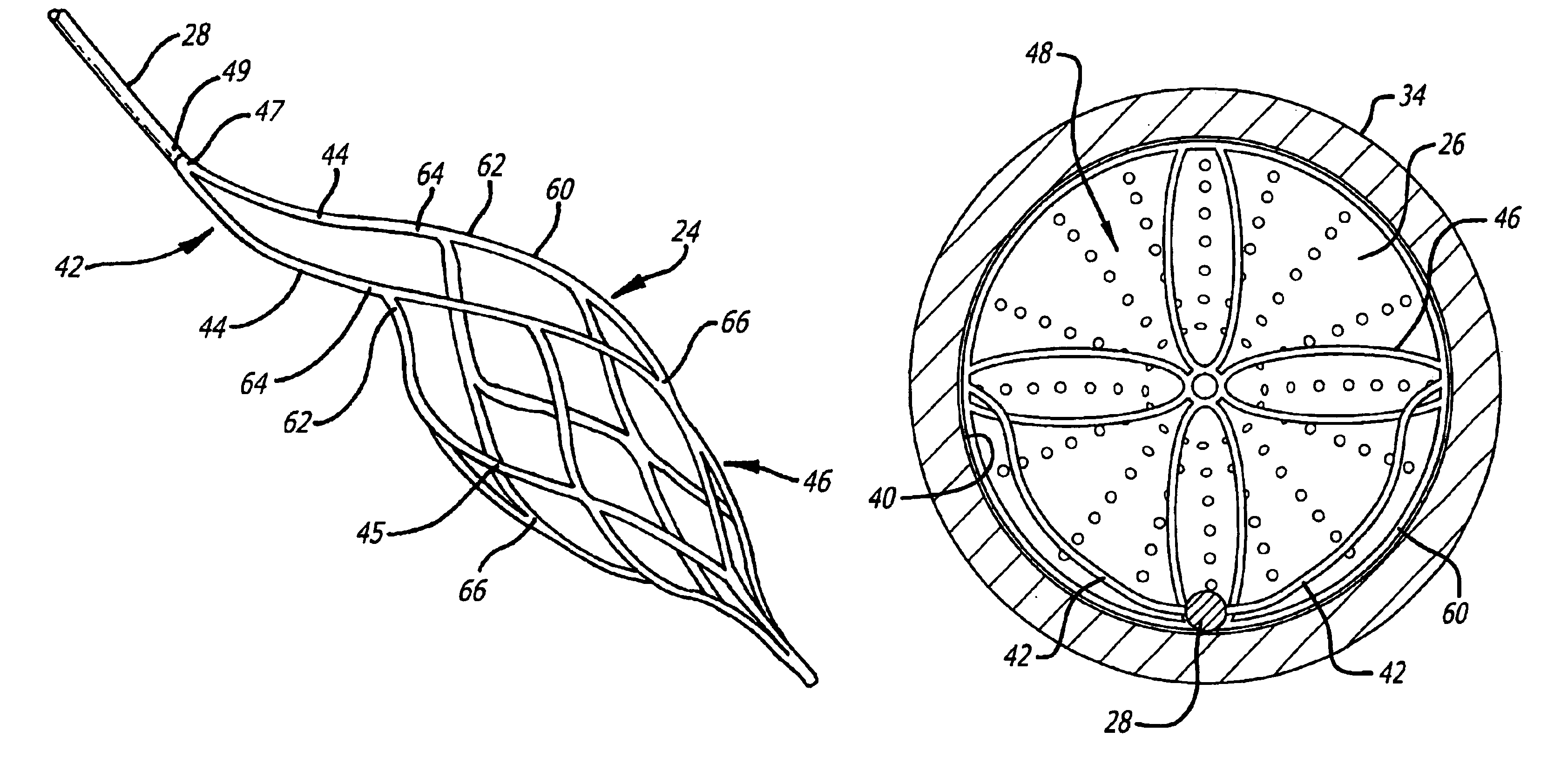
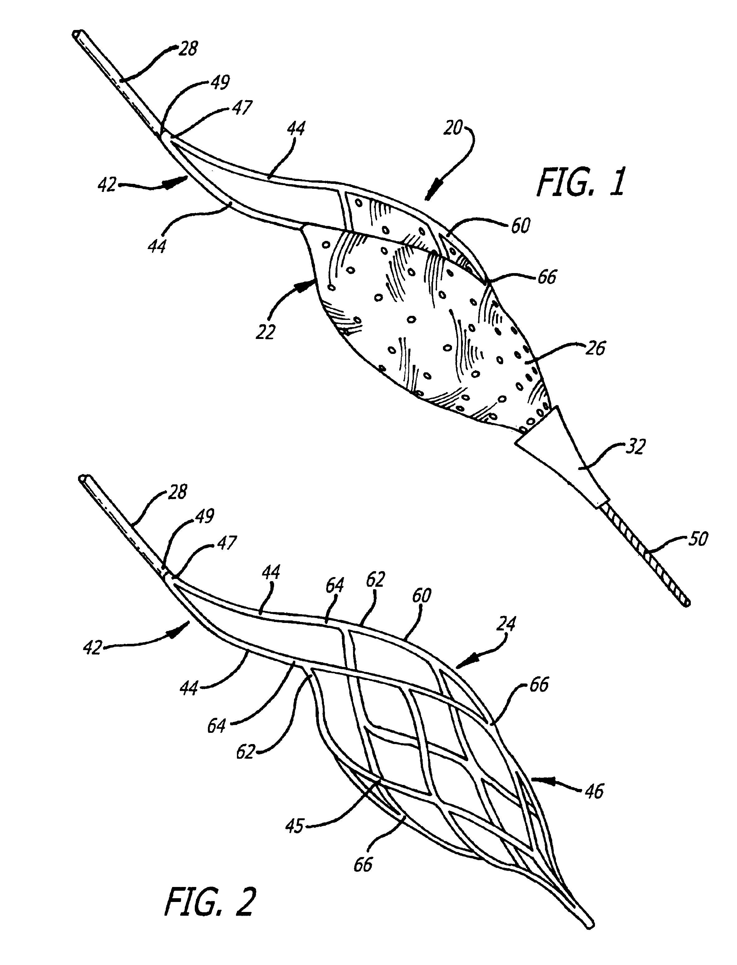
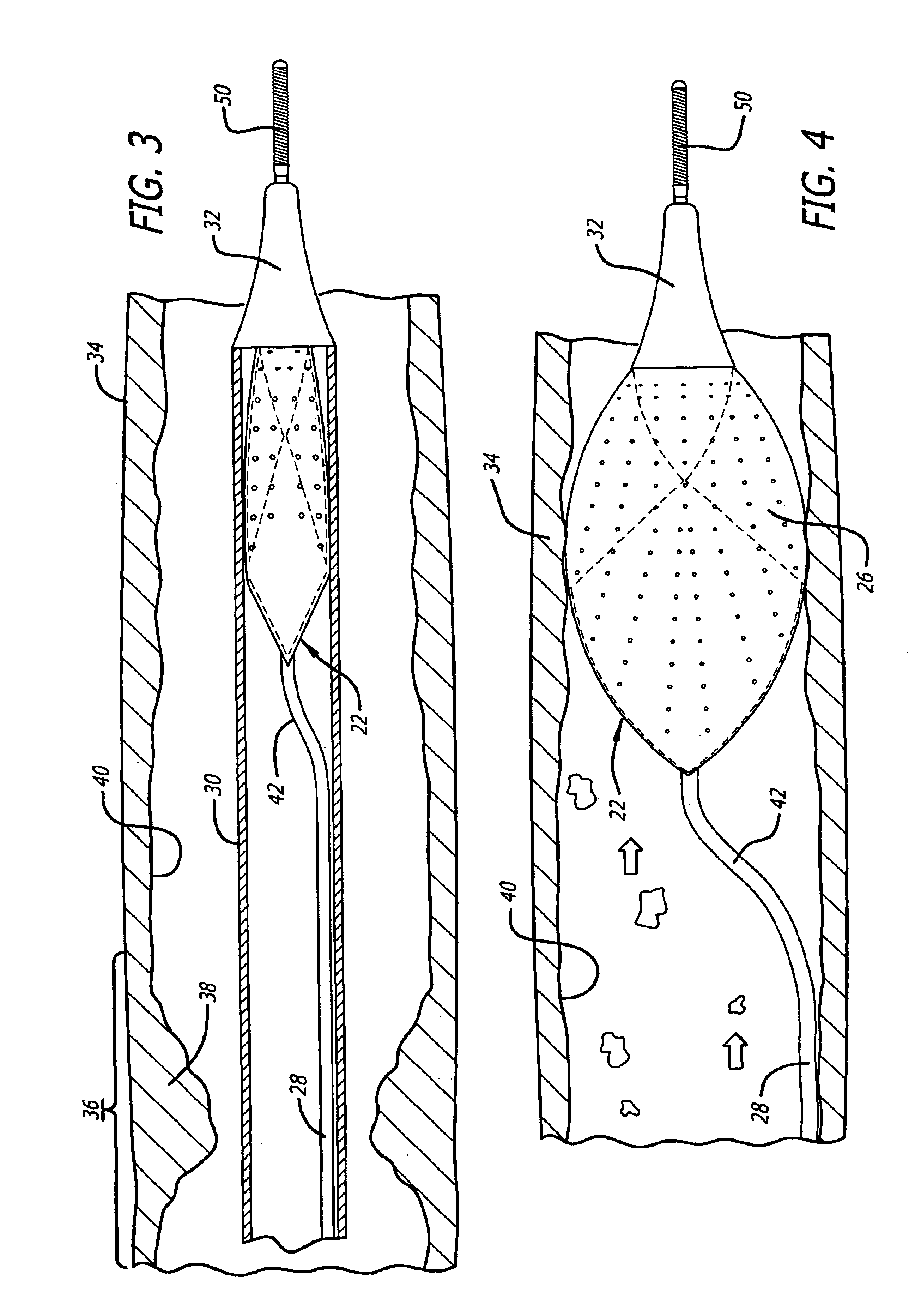
[0033]Turning now to the drawings, in which like reference numerals represent like or corresponding elements in the drawings, FIGS. 1 and 2 illustrate one particular embodiment of an embolic filtering device 20 incorporating features of the present invention. This embolic filtering device 20 is designed to capture embolic debris which may be created and released into a body vessel during an interventional procedure. The embolic filtering device 20 includes an expandable filter assembly 22 having a self-expanding basket or cage 24 and a filter element 26 attached thereto. In this particular embodiment, the expandable filter assembly 22 is mounted on the distal end of an elongated tubular shaft, such as a guide wire 28. A restraining or delivery sheath 30 (FIG. 3) extends coaxially along the guide wire 28 to maintain the expandable filter assembly 22 in its unexpanded position until it is ready to be deployed within the patient's vasculature. The expandable filter assembly 22 is deplo...


PUM


Abstract

An expandable cage used in conjunction with an embolic filtering device has a strut configuration including a proximal strut assembly coupled to a distal strut assembly. A filter can be attached to the distal strut assembly which has an inlet opening. The proximal strut assembly is “offset” from the distal strut assembly in that these proximal struts extend substantially along the vessel wall of the patient, rather than being “centered” in the body vessel when the cage is expanded in a body vessel. As a result, there is little cage structure directly in front of the opening of the filter, resulting in a virtually unobstructed opening for the filter.

Description

BACKGROUND OF THE INVENTION[0001]The present invention relates generally to filtering devices used when an interventional procedure is being performed in a stenosed or occluded region of a body vessel to capture embolic material that may be created and released into the vessel during the procedure. The present invention is more particularly directed to an embolic filtering device made with an expandable cage or basket having good flexibility and bendability.[0002]Numerous procedures have been developed for treating occluded blood vessels to allow blood to flow without obstruction. Such procedures usually involve the percutaneous introduction of an interventional device into the lumen of the artery, usually by a catheter. One widely known and medically accepted procedure is balloon angioplasty in which an inflatable balloon is introduced within the stenosed region of the blood vessel to dilate the occluded vessel. The balloon dilatation catheter is initially inserted into the patient...

Claims


Application Information

Patent Timeline
Patent Type & Authority Patents(United States)
IPC IPC(8): A61F2/01
CPCA61F2/013B82Y20/00B82Y30/00A61F2002/018A61F2230/0076A61F2230/008A61F2230/0006A61F2230/005
Inventor BOYLE, WILLIAM J.PAPP, JOHN E.
Owner ABBOTT CARDIOVASCULAR
Why Eureka
Try Eureka