

Method for slice position planning of tomographic measurements, using statistical images
a tomographic and statistical image technology, applied in tomography, instruments, applications, etc., can solve problems such as difficulty in comparing images obtained from respective scans
- Summary
- Abstract
- Description
- Claims
- Application Information
AI Technical Summary
Benefits of technology
Problems solved by technology
Method used
Image

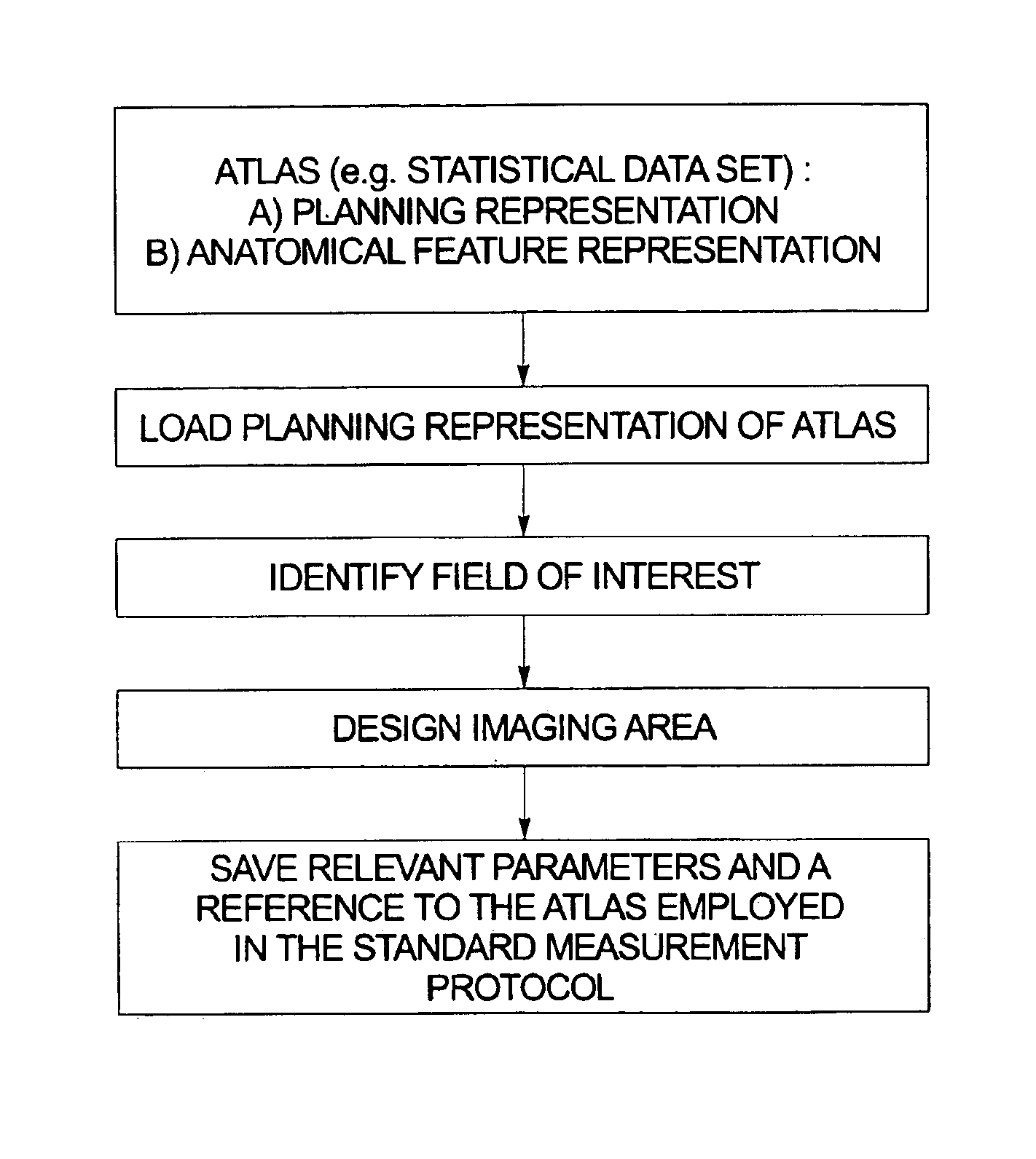
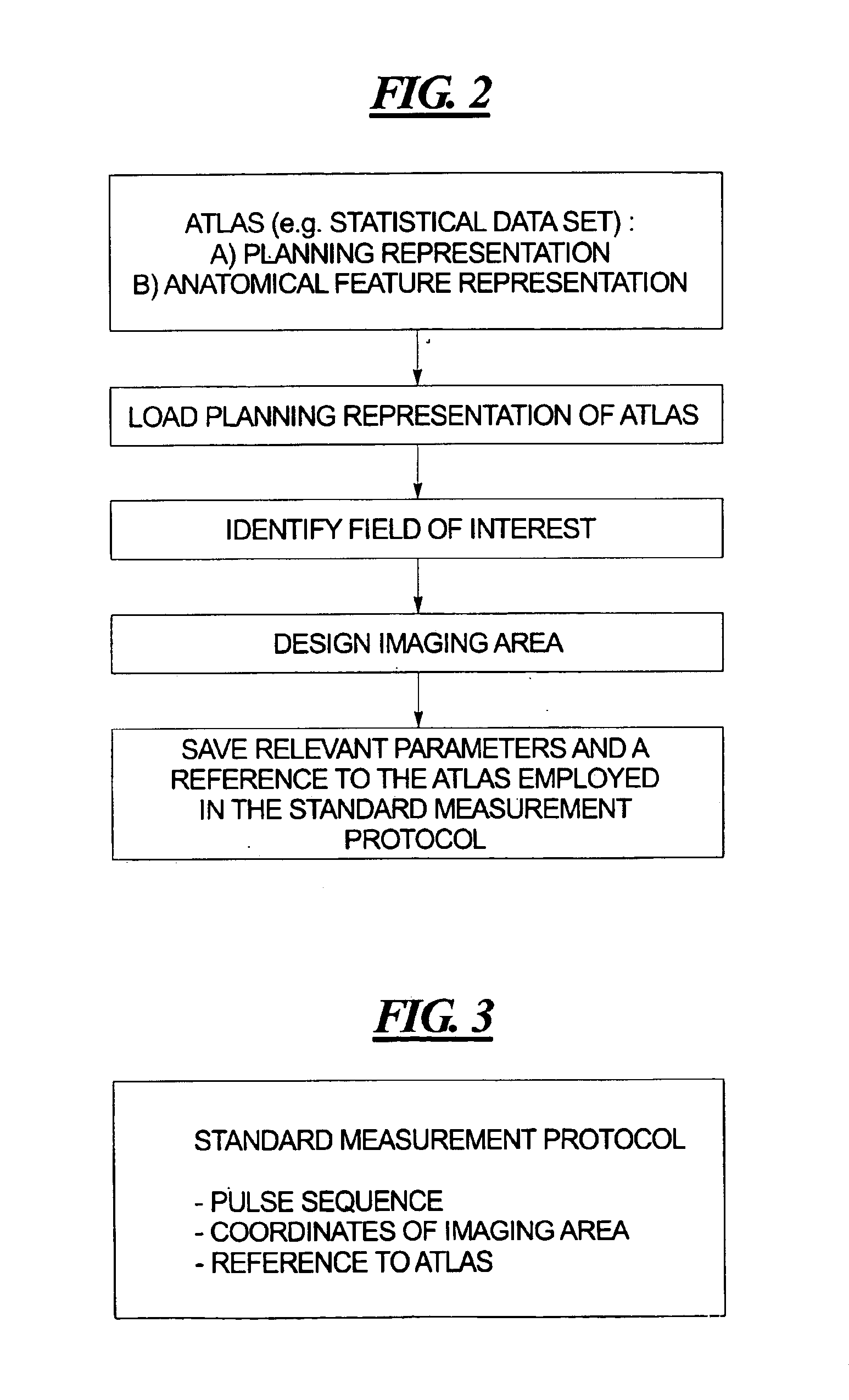
Examples
Embodiment Construction
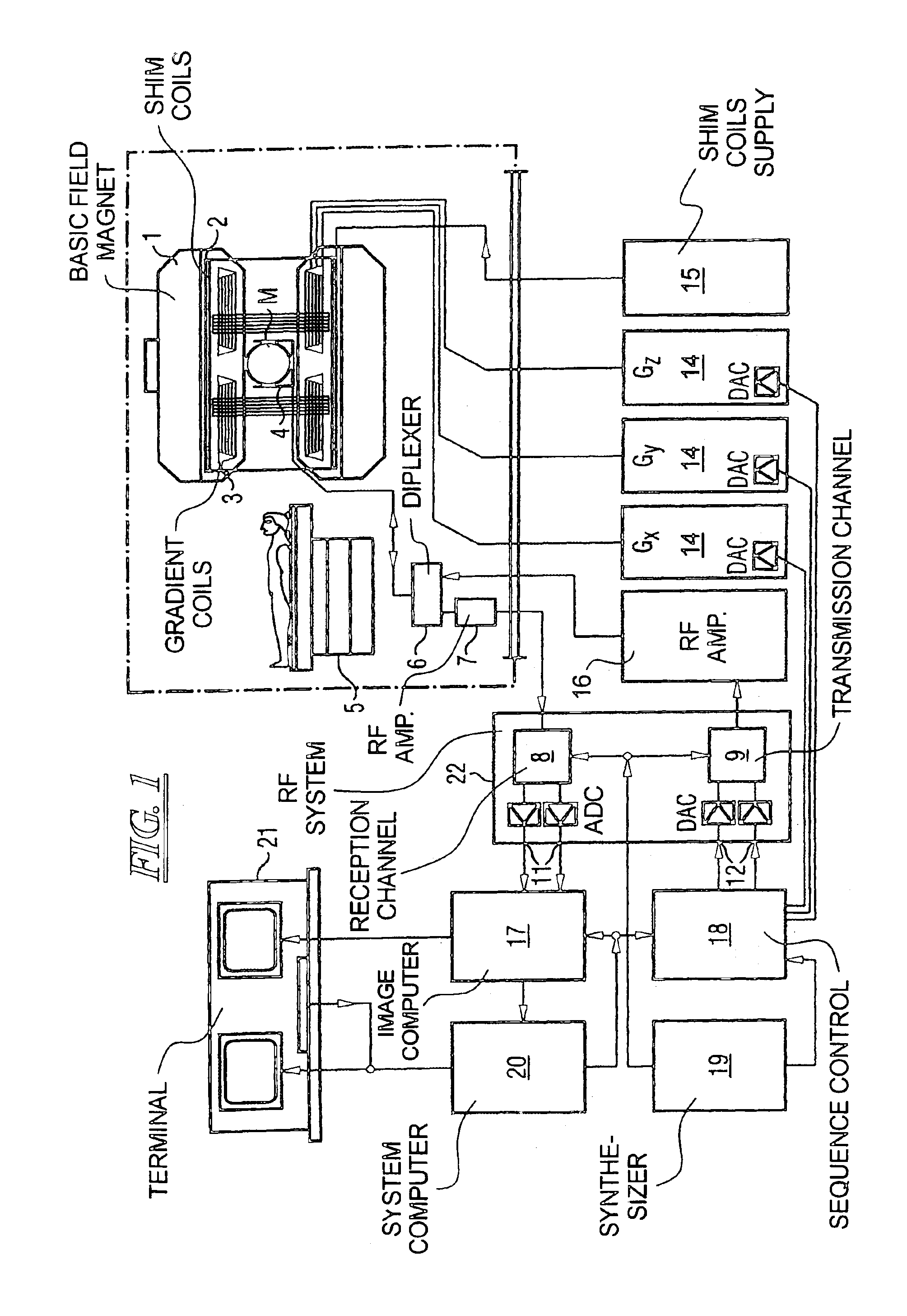
[0027]FIG. 1 schematically illustrates a magnetic resonance imaging (tomography) apparatus for generating a magnetic resonance image of a subject, as an example of a tomographic imaging modality operable according to the present invention. The components of the nuclear magnetic resonance tomography apparatus correspond to those of a conventional tomography apparatus, but it is controlled according to the invention. A basic field magnet 1 generates a time-constant, intense magnetic field for polarization (alignment) of the nuclear spins in the examination region of a subject such as, for example, a part of a human body to be examined. The high homogeneity of the basic magnetic field required for the nuclear magnetic resonance measurement is defined in a spherical measurement volume M in which the part of the human body to be examined is introduced. For supporting the homogeneity demands and, in particular, for eliminating time-invariable influences, shim plates of ferromagnetic mater...
PUM
 Abstract
Description
Claims
Application Information