Multi-modal optical tissue diagnostic system

a tissue diagnostic and optical tissue technology, applied in the direction of fluorescence/phosphorescence, therapy, diagnostics using spectroscopy, etc., can solve the problems of insufficient assessment of tissue characteristics, insufficient absorption spectroscopy alone, and insufficient morphological changes due to disease, etc., to achieve accurate diagnosis of tissue condition and accurate measurement of tissue characteristics. , the effect of accurate tissue diagnosis

Inactive Publication Date: 2005-12-13
SPECTRX
View PDF234 Cites 94 Cited by
  • Summary
  • Abstract
  • Description
  • Claims
  • Application Information

AI Technical Summary

Benefits of technology

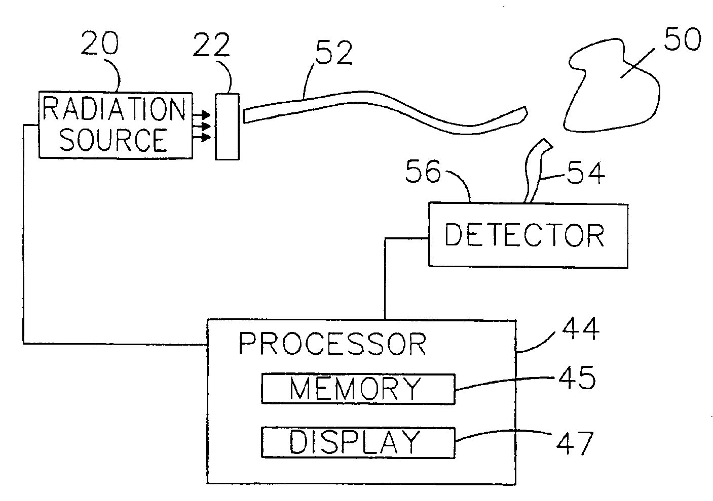
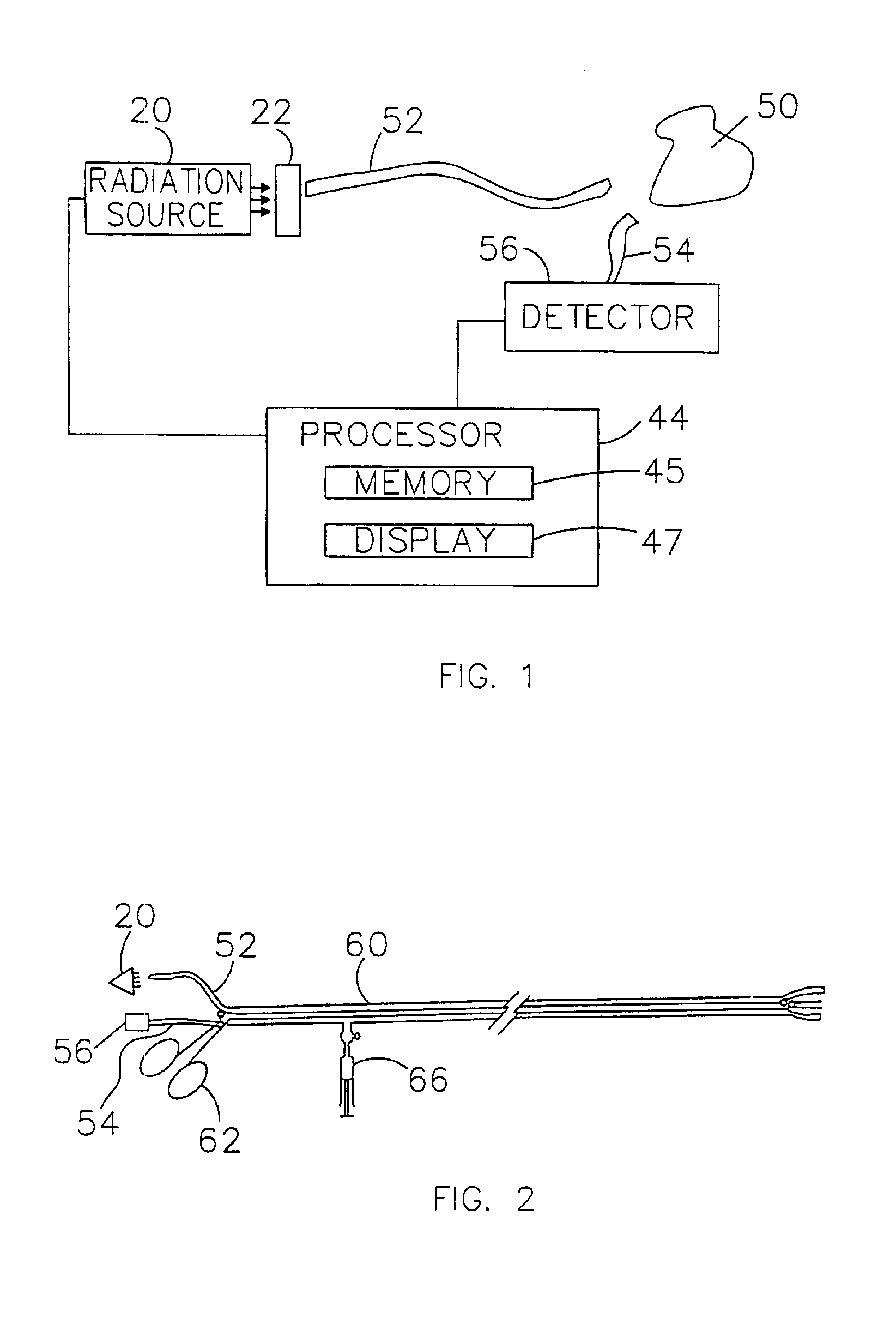
[0015]The invention focuses on providing methods and apparatus that provide accurate measurements of changes in characteristics of tissues. The methods and apparatus according to the invention combine more than one optical modality (spectroscopic method), including but not limited to fluorescence, absorption, reflectance, polarization anisotropy, and phase modulation to decouple morphological and biochemical changes associated with tissue changes, and thus to prov

Problems solved by technology

None of the prior art methods discussed above alone is sufficient to accurately measure changes in tissue characteristics.
Likewise, absorption spectroscopy alone is insufficient to assess changes in tissue characteristics because it is less sensitive to biochemical changes in tissue, as compared to fluorescence spectrosco

Method used


Image

  • Multi-modal optical tissue diagnostic system
  • Multi-modal optical tissue diagnostic system
  • Multi-modal optical tissue diagnostic system

Examples


Embodiment Construction

[0037]In the prior art methods in the Background of the Invention Section, the information content of the interaction of light (and consequently the spectroscopic method used) is, generally speaking, specific to the type of change in tissue. That is, tumorous tissue differs from normal tissue in several ways. Tumorous tissue is generally derived from normal tissue after the latter has undergone several changes. These changes can be induced by various intrinsic and extrinsic factors. These include the presence of certain inherited traits, chromosomal mutation, virus induced malignant transformation of cells and the mutagenic effects of UV and X-ray irradiation, to name a few.

[0038]The earliest changes that occur in the course of normal cells becoming malignant are biochemical. One of the first changes noted is that of increased glycolytic activity which allows tumors to grow to a large size with decreased oxygen requirements. Invasive tumor cells secrete type IV collegenase destroyin...


PUM


Abstract

An apparatus and method according to the invention combine more than one optical modality (spectroscopic method), including but not limited to fluorescence, absorption, reflectance, polarization anisotropy, and phase modulation, to decouple morphological and biochemical changes associated with tissue changes due to disease, and thus to provide an accurate diagnosis of the tissue condition.

Description

[0001]This application is a Continuation of application Ser. No. 09 / 786,781 filed Mar. 9, 2001 now abandoned which is a 371 of PCT / US99 / 20646, filed Sep. 10, 1999, which published on Mar. 23, 2000 with Publication No. WO 00 / 15101 in the English language and which claimed priority to U.S. Provisional Application Nos. 60 / 099,875 filed Sep. 11, 1998 and 60 / 143,579 filed Jul. 13, 1999.BACKGROUND OF THE INVENTION[0002]1. Field of the Invention[0003]The invention relates to apparatus and methods for determining tissue characteristics of, for example, a human or animal.[0004]2. Background of the Related Art[0005]Spectroscopic methods for determining tissue characteristics are known and have been widely used to interrogate changes in tissue. A number of these distinct spectroscopic techniques are available that provide specific information depending on the nature of the interaction of light with cells and the natural chromophores present in tissue. These interactions include the absorption ...

Claims


Application Information

Patent Timeline
IPC IPC(8): A61B1/00G01N21/64A61B1/05A61B5/00A61B10/00G01N21/27G01N21/33
CPCA61B5/0071A61B5/0075A61B5/0084A61B1/043A61B5/7264
Inventor FAUPEL, MARK L.BAMBOT, SHABBIR B.HARRELL, TIMAGRAWAL, ANANT
Owner SPECTRX
Why Eureka
Try Eureka