Assays for autoantibodies

a technology of autoantibodies and autoantibody, which is applied in the field of autoantibodies assays, can solve the problems of affecting the inability to carry out current assays in specially equipped laboratories, and the distortion of the feedback system, so as to achieve the effect of improving the accuracy of autoantibodies and reducing the risk of autoantibodies

Inactive Publication Date: 2006-02-21
R S R
View PDF26 Cites 12 Cited by
  • Summary
  • Abstract
  • Description
  • Claims
  • Application Information

AI Technical Summary

Problems solved by technology

In thyroid disease, however, the above feedback system is often distorted.
Tg levels are usually measured by assays which depend on monoclonal and / or polyclonal antibodies to Tg but if autoantibodies to Tg are present in patient test samples, these autoantibodies can interfere with the Tg assays, giving erroneous results.
However, there have been some major limitations associated with current assay methods for autoantibodies, for example:—the current assays can only be carried out away from the patient in specially equipped laboratories; andthe current assays can only be carried out by experienced personnel and take several hours to complete.

Method used


Image

  • Assays for autoantibodies
  • Assays for autoantibodies
  • Assays for autoantibodies

Examples


example 1

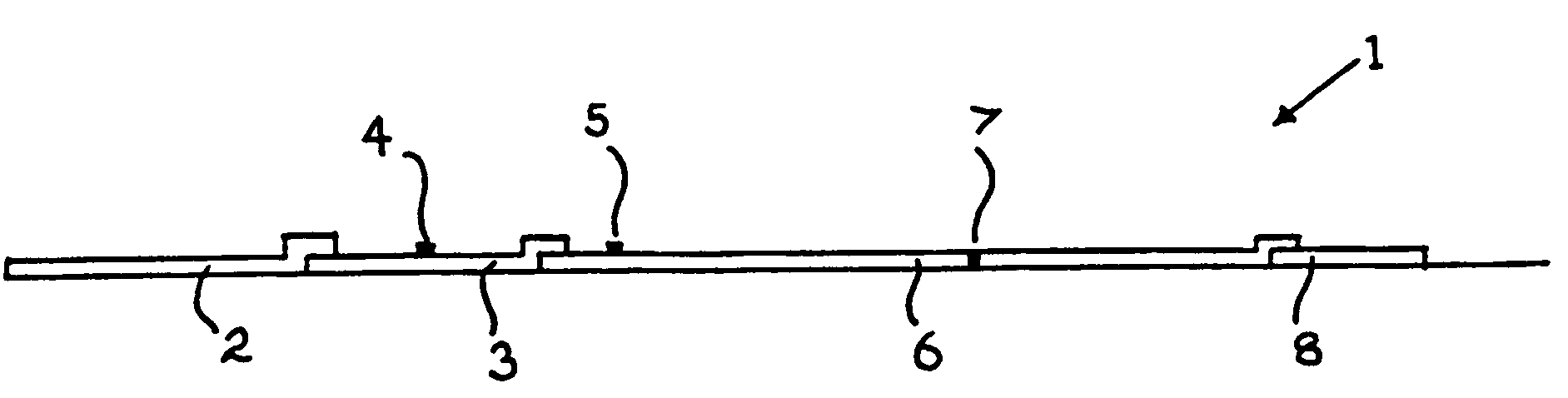
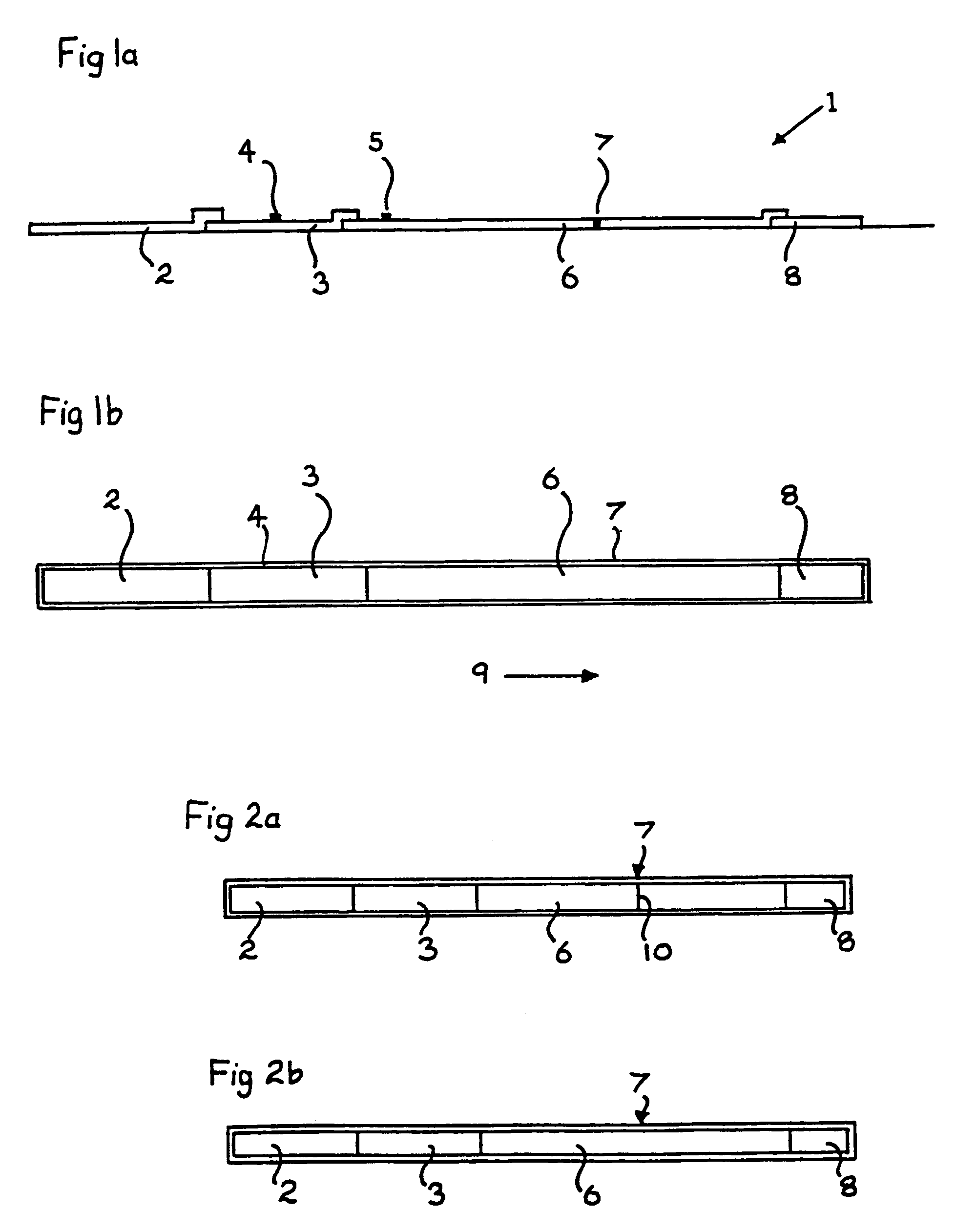
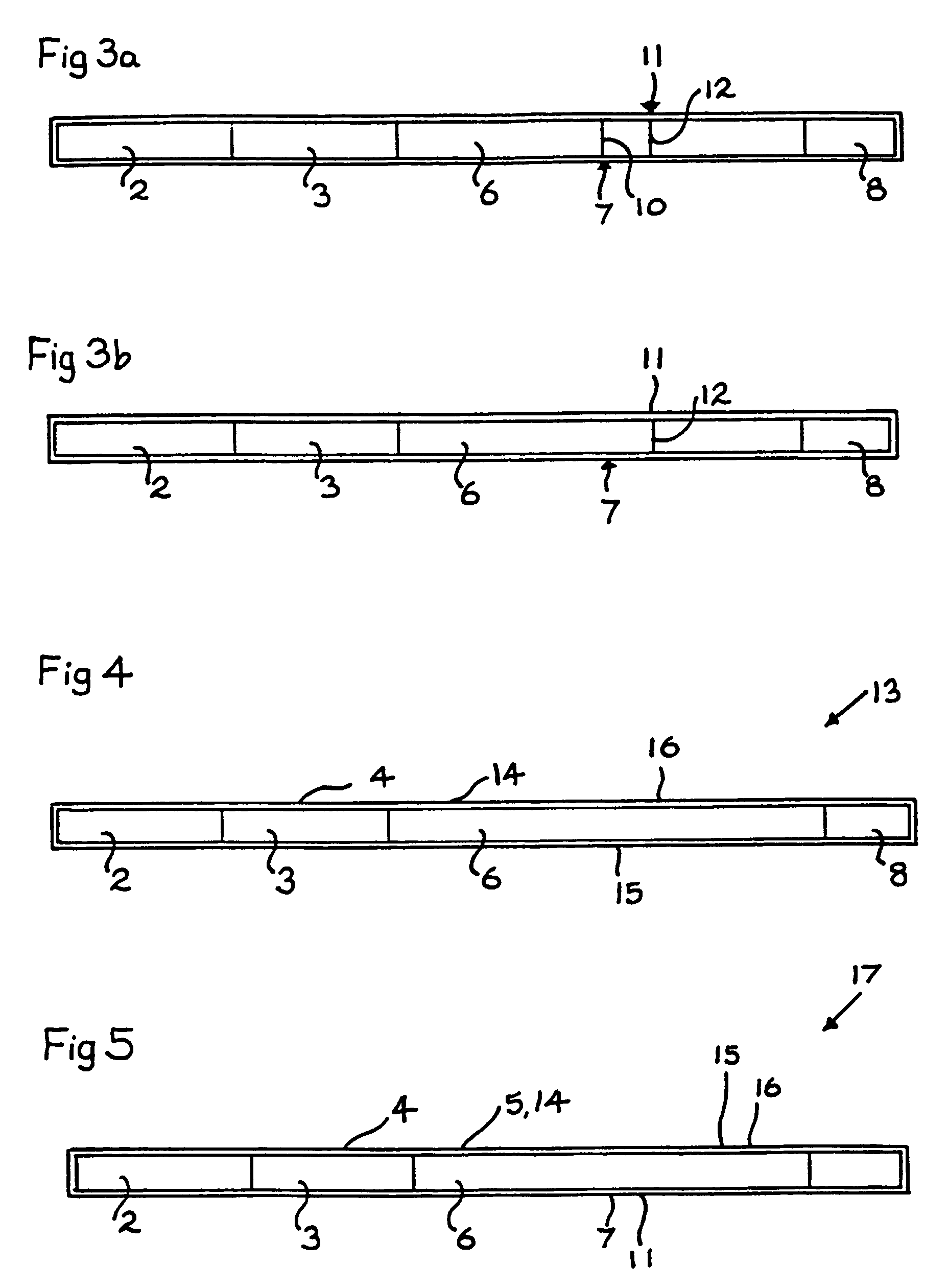
[0154]The following tables give the results obtained using a TPO screening kit as shown in FIGS. 1a and 1b and a Tg screening kit as shown in FIG. 4.

[0155]

TABLE 1Patient sample results - TPOAbTPOAb rapid assayTPOAbconcentration byQualitativeradioactive assayresult by rapidPatient sample(U / ml)assay+TPOAb1165.6(+)+219.0(+)+314.6(+)+456.6(+)+550.1(+)+6108.7(+)+7171.4(+)+890.7(+)+−TPOAb90.2(−)−100.1(−)−110.2(−)−120.2(−)−130.3(−)−140.3(−)−

[0156]

TABLE 2Patient sample results - TgAbTgAb rapid assayTgAbconcentration byQualitativeradioactive assayresult by rapidPatient sample(U / ml)assay+TgAb152.4 (+)+245.3 (+)+−TgAb3Neg−4Neg−

example 2

[0157]This Example describes screening for autoantibodies to Tg using a kit as illustrated in FIGS. 7 and 8.

[0158]90 μl of plasma (or sera) or 30 μl of whole blood plus 60 μl of a diluent buffer (150 mM NaCl, 20 mM Tris pH7.6) were used. Results were obtained after 10 minutes. Prior art reference radioactive method was based on that of Beever et al Clinical Chemistry 35 (1989) 1949–954.

[0159]

TABLE 3(a) Results obtained in whole blood or plasma obtainedfrom 30 healthy blood donors.whole blood(Reference methodplasmacannot be used with(n = 30)whole blood)Tg autoantibody positive by 3 / 30—reference radioactive testTg autoantibody positive by 3 / 30 1 / 30a method according to thepresent invention employinga kit as illustrated inFIGS. 7 and 8Tg autoantibody negative by27 / 30—reference radioactive testTg autoantibody negative27 / 3029 / 30by a method according tothe present inventionemploying a kit asillustrated in FIGS. 7and 8

[0160]

TABLE 4(b) Results obtained with sera from patients withsystemic l...

example 3

[0162]The Example describes screening for autoantibodies to TPO using a kit as illustrated in FIGS. 9 and 10. 90μl of plasma (or sera) or 30 μl of whole blood plus 60 μl of a diluent buffer (150 mM Nacl; 20 mM Tris pH 7.6) were used. Results were obtained after 10 minutes. Prior art reference radioactive method was based on that of Beever et al Clinical Chemistry 35 (1989) 1949–1954.

[0163]

TABLE 5(a) Results obtained in whole blood or plasma obtainedfrom 30 healthy blood donorswhole blood(reference methodcannot be used withplasmawhole blood)TPO autoantibody positive 3 / 30—by reference radioactivetestTPO autoantibody positive 3 / 30 3 / 30by a method according tothe present inventionemploying a kit asillustrated in FIGS. 9and 10TPO autoantibody negative27 / 30—by reference radioactivetestTPO autoantibody negative27 / 3027 / 30by a method according tothe present inventionemploying a kit asillustrated in FIGS. 9and 10

[0164]

TABLE 6(b) Results obtained in sera from patients with suspectedautoimmune ...


PUM

PropertyMeasurementUnit
pHaaaaaaaaaa
colorimetric changeaaaaaaaaaa
Tgaaaaaaaaaa

Abstract

A method and kit for screening a sample of body fluid for at least one autoantibody to at least one antigen. A source of at least one antigen to the autoantibody is provided. A substrate having immobilized thereto at least one antibody to the antigen is also provided. The antigen source is contacted with the sample of body fluid, so as to obtain a mixture wherein the antigen is allowed to substantially bind with the autoantibody, when the latter is present in the sample of body fluid. The mixture is allowed to flow relative to the substrate so as to allow the mixture to contact the antibody immobilized to the substrate. Labeling means are provided to permit monitoring of binding of the autoanitbody and the antigen present in the mixture, so as to provide an indication of the presence of the autoanitbody in the sample of body fluid.

Description

CROSS REFERENCE TO RELATED APPLICATIONS[0001]This application is a National Phase application concerning a filing under 35 U.S.C. 371, of International Patent Application Serial No. PCT / GB99 / 03548, filed Oct. 27, 1999, which claims priority on British Patent Application Serial No. 9823397.6, filed Oct. 27, 1998, both of which are incorporated herein by reference.BACKGROUND OF THE INVENTION[0002]1. Field of the Invention[0003]The present invention is concerned with assays for screening a sample of body fluid for autoantibodies to various antigens. In particular, the present invention is concerned with screening a sample of body fluid for autoantibodies associated with autoimmune thyroid disease.[0004]2. Description of Related Art[0005]Autoimmune diseases are characterised by the presence of circulating autoantibodies and in autoimmune thyroid disease, for example, the autoantibodies are directed to three different thyroid proteins, namely thyroid peroxidase (TPO), thyroglobulin (Tg) ...

Claims


Application Information

Patent Timeline
Patent Type & Authority Patents(United States)
IPC IPC(8): G01N33/543A61K45/00A61P37/06G01N33/53G01N33/548G01N33/558G01N33/564G01N33/566G01N33/76
CPCG01N33/558G01N33/564G01N33/76G01N2800/24Y10S435/81Y10S435/971Y10S436/824Y10S436/815Y10S435/973Y10S436/805Y10S435/975A61P37/06G01N33/54388
Inventor BURNE, PETER JOHNSMITH, BERNARD REES
Owner R S R
Why Eureka
Try Eureka