
Segmentation of 3D medical structures using robust ray propagation

Inactive Publication Date: 2006-03-21
SIEMENS MEDICAL SOLUTIONS USA INC
View PDF3 Cites 57 Cited by
  • Summary
  • Abstract
  • Description
  • Claims
  • Application Information

AI Technical Summary

Benefits of technology

[0007]A robust and efficient method for segmentation of 3D structures in CT and MR images is provided. The method is based on 3D ray propagation by mean-shift analysis with a smoothness constraint. Ray propagation is used to guide an evolving surface due to its computational efficiency. In addition, non-parametric analysis and shape priors are incorporated to the method for robust convergence.

Problems solved by technology

The segmentation of three-dimensional (3D) structures in CT (computerized tomography) and MR (magnetic resonance) images is an inherently difficult and time consuming problem.
In addition, the processing in 3D space implies an increased computational complexity, which makes impractical the 3D extension of most two-dimensional (2D) segmentation algorithms.
However, such initialization of deformable models is typically a difficult task because a 3D model must be created inside 3D data by using a 2D display device.
In addition, the current deformable model approaches are computationally expensive.
This is due to the additional dimension in the case of level set methods, or expensive reinitialization of parameters in the case of explicit surface evolution techniques.

Method used


Image

  • Segmentation of 3D medical structures using robust ray propagation
  • Segmentation of 3D medical structures using robust ray propagation
  • Segmentation of 3D medical structures using robust ray propagation

Examples


Embodiment Construction

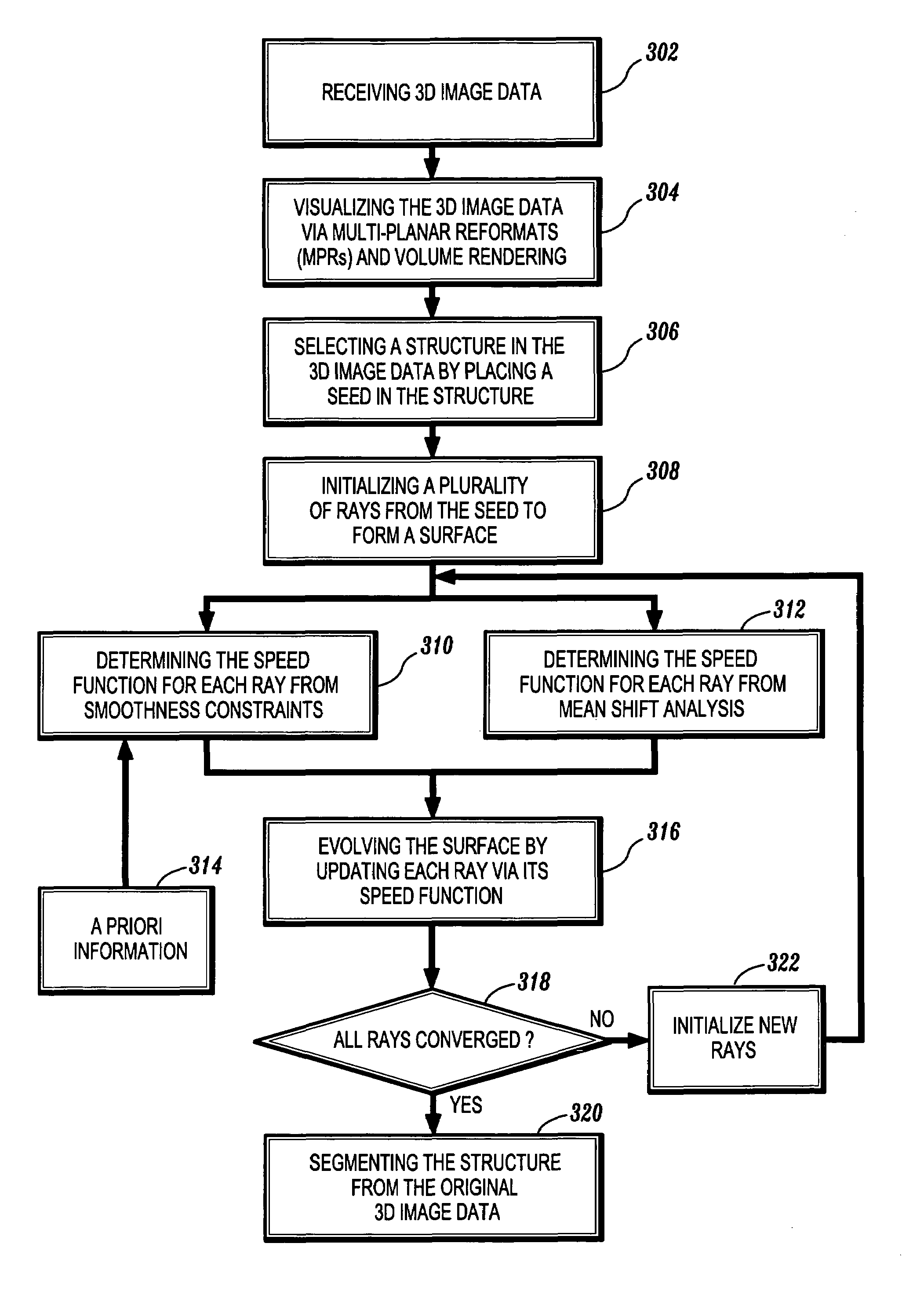
[0017]Preferred embodiments of the present invention will be described hereinbelow with reference to the accompanying drawings. In the following description, well-known functions or constructions are not described in detail to avoid obscuring the invention in unnecessary detail.

[0018]The present invention provides an efficient, robust, and user-friendly method for the interactive segmentation of 3D medical structures, e.g., aneurysms, from contrast enhanced CT or MR data. The primary innovation of the method is the boundary propagation by mean-shift analysis combined with a smoothness constraint. As a result, the method of the present invention is robust to both (1) outliers in the data and (2) missing data structures, e.g., surfaces that are not well defined or missing. The first property is guaranteed by a mean-shift procedure, while the second is obtained through the use of a priori information on boundary smoothness. Furthermore, due to the method's computationally efficient fra...


PUM


Abstract

A method for segmentation of 3D structures in CT and MR images is provided. The method is based on 3D ray propagation by mean-shift analysis with a smoothness constraint. Ray propagation is used to guide an evolving surface due to its computational efficiency and shape priors are incorporated for robust convergence. The method includes the steps of receiving 3D image data; visualizing the 3D image data on a display device; selecting a structure in the 3D image data by placing a seed in the structure; initializing a plurality of rays from the seed to form a surface; determining a speed function of each of the rays; evolving the surface by propagating the rays based on the speed function of each of the rays; converging the rays on a boundary of the structure; and segmenting the structure when all of the rays have converged on the structure's boundary.

Description

CROSS REFERENCE TO RELATED APPLICATION[0001]The present application claims priority to U.S. Provisional Patent Application Ser. No. 60 / 373,684, filed Apr. 18, 2002, which is hereby incorporated by reference.BACKGROUND OF THE INVENTION[0002]1. Field of the Invention[0003]The present invention relates generally to computer vision and imaging systems, and more particularly, to a system and method for segmentation of 3D medical structures, e.g., organs, aneurysms and brain tumors, using robust ray propagation.[0004]2. Description of the Related Art[0005]The segmentation of three-dimensional (3D) structures in CT (computerized tomography) and MR (magnetic resonance) images is an inherently difficult and time consuming problem. The accurate localization of object surfaces is influenced by the presence of significant noise levels, generated through partial-volume effects and by image acquisition devices. In addition, the processing in 3D space implies an increased computational complexity,...

Claims


Application Information

Patent Timeline
IPC IPC(8): G06T17/20G06T5/00G06T17/00
CPCG06T7/0083G06T7/0089G06T17/00G06T2207/30016G06T2207/10088G06T2207/20101G06T2207/20168G06T2207/10081G06T7/12G06T7/149
Inventor TEK, HUSEYINCOMANICIU, DORINWILLIAMS, JAMES P.
Owner SIEMENS MEDICAL SOLUTIONS USA INC
Learn More