System and method for fusion-aligned reprojection of incomplete data

a technology of incomplete data and fusion alignment, applied in the field of radiological and radiation therapy, can solve the problems of inaccurate targeting, irregular white border, and inability to accurately target the data set obtained on the day of treatment to be used for preparing the patient model, etc., to achieve accurate targeting, accurate radiation application, and reduced damage to healthy tissue

Inactive Publication Date: 2006-05-16
TOMOTHERAPY INC
View PDF21 Cites 67 Cited by
  • Summary
  • Abstract
  • Description
  • Claims
  • Application Information

AI Technical Summary

Benefits of technology

[0015]The method of the present invention is advantageous in that the availability of only one limited data sinogram / image will not affect the ability to perform accurate delivery verification, dose reconstruction, patient set-up or the like. The limited data image or “first image” is fused to a previously taken complete image or “second image.” The sinogram representing the fused image is compared to the limited data sinogram, and the augmented limited data sinogram is prepared therefrom. From the augmented limited data sinogram the fusion-aligned reprojected (FAR) image is obtained. The FAR image is used to accurately apply radiation to the treatment area, which may be positioned differently than as shown in the previously obtained complete image.
[0016]The advantages of obtaining current data at the time of treatment or even dosage verification are many. Damage to healthy tissue will be reduced, and the cancerous or diseased tissue will be more accurately targeted. These differences are especially critical in areas that have frequent internal anatomy changes, such as the torso or prostate.

Problems solved by technology

Uncertainty in the positioning of the patient with respect to the original CT image can undermine the conformality of the radiation delivery.
Unfortunately, the data sets obtained on the day of treatment to be used for preparing the patient model are often incomplete.
Not only is there a limited field of view, the data around the edges contains significant artifacts so that the image has an irregular white border and internal values are distorted.
There may be other limitations that result in the collection of incomplete data sets.
This has been done by adding together sinogram data, but requires that the imaging apparatus or patient position can be reliably modified accordingly, which is not always possible.
Further, the problem of developing artifacts is still present due to the significant degree of mismatch between such data sets, and the additional handling of the patient is more costly, time intensive and can be difficult for frail patients.
Reconstruction of incomplete data sets using available techniques results in images that do not show the complete extent of the patient's body, can have artifacts and incorrect voxel values, and thus, limit the extent to which the images can be used for delivery verification, dose reconstruction and patient set-up, deformable patient registration and deformable dose registration.

Method used


Image

  • System and method for fusion-aligned reprojection of incomplete data
  • System and method for fusion-aligned reprojection of incomplete data
  • System and method for fusion-aligned reprojection of incomplete data

Examples


Embodiment Construction

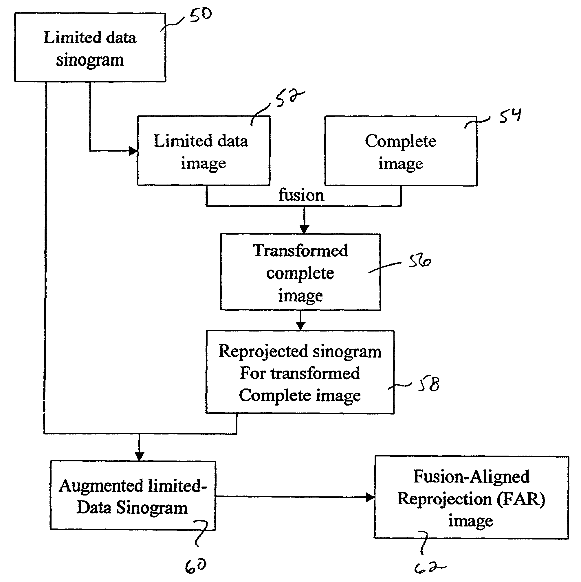
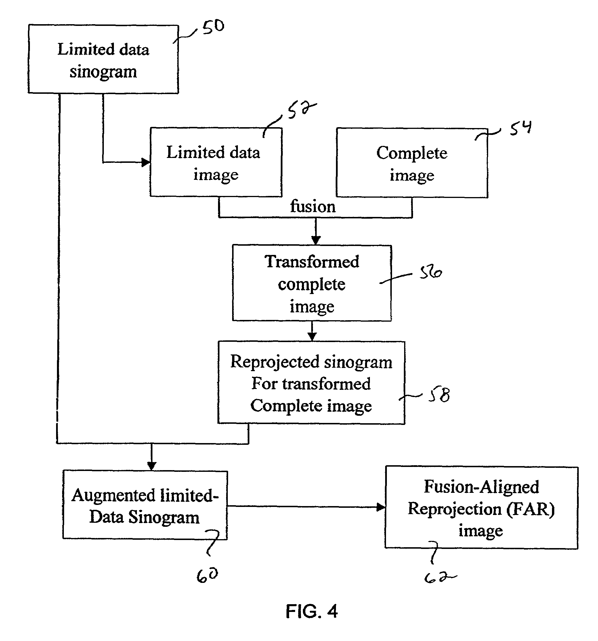
[0029]A preferred method in accordance with the present invention is shown in the flowchart of FIG. 4. A limited data sinogram 50 representing the treatment area is obtained from a patient. In one preferred embodiment of the present invention, the limited data sinogram 50 is prepared near the time that the patient is receiving his or her radiation treatment. However, the limited data sinogram 50 may be obtained at any time.

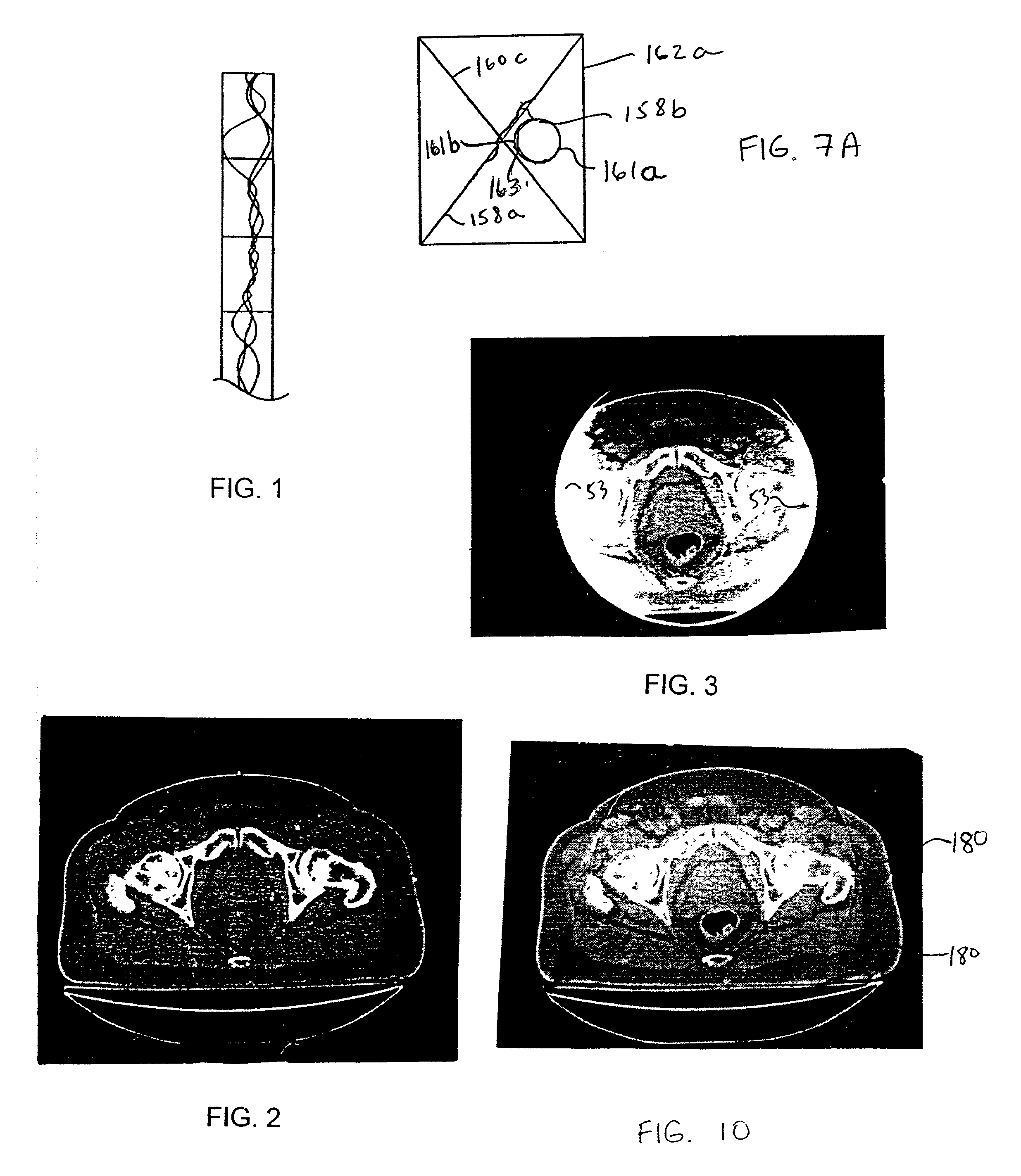
[0030]The limited data sinogram 50 is reconstructed to a limited data image 52, as seen in the example of FIG. 3, and represented schematically in FIG. 6 as limited object 156. FIG. 3 contains a significant amount of artifacts such as the white irregular border 53, and some distortion of image values. By way of example, the treatment area targeted in FIG. 3 is a prostate gland. The method can be applied to images of any part of the body, or be used in veterinary or radiological applications.

[0031]A complete image 54 of the same patient and same treatment area is s...


PUM


Abstract

The present invention provides a system and method of using current but incomplete data to prepare an approximated complete image of a patient potentially undergoing radiation therapy. A limited patient image, such as that obtained from a CT scan is fused with a complete image of the same area using image registration techniques. The fused image is converted to sinogram data. This data is compared to sinogram data corresponding to the limited patient image to determine what data exists beyond the scope of the limited sinogram. Any additional data is added to the limited data sinogram to obtain a complete sinogram. This is reconstructed into an image that approximates the complete image that would have been taken at the time the limited image was obtained.

Description

FIELD OF THE INVENTION[0001]This invention relates generally to radiation therapy and radiology, and more particularly to a method for reconstructing incomplete patient data for radiation therapy set-up and treatment verification.BACKGROUND OF THE INVENTION[0002]Medical equipment for radiation therapy treats tumorous tissue with high energy radiation. The amount of radiation and its placement must be accurately controlled to ensure both that the tumor receives sufficient radiation to be destroyed, and that the damage to the surrounding and adjacent non-tumorous tissue is minimized.[0003]In external source radiation therapy, a radiation source external to the patient treats internal tumors. The external source is normally collimated to direct a beam only to the tumorous site. The source of high energy radiation may be x-rays, or electrons from linear accelerators in the range of 2–25 MeV, or gamma rays from highly focused radioisotopes such as a Co.sup.60 source having an energy of 1...

Claims


Application Information

Patent Timeline
Patent Type & Authority Patents(United States)
IPC IPC(8): G06K9/00G06K9/32G06K9/36G06K9/40G06K9/46A61B6/03A61B6/08A61N5/10G06F19/00G06T1/00G06T11/00
CPCA61B6/08A61B6/5235G06T11/005A61B6/508A61N5/1049
Inventor RUCHALA, KENNETH J.OLIVERA, GUSTAVO H.MACKIE, THOMAS R.KAPATOES, JEFFREY M.RECKWERDT, PAUL J.
Owner TOMOTHERAPY INC
Why Eureka
Try Eureka