
Detection of ribcage boundary from digital chest image

a digital chest image and ribcage boundary technology, applied in the field of image processing technique, can solve the problems of reducing the accuracy of computer-aided diagnosis as a whole, reducing the above-described conventional automatic detection technique tends to fail in the detection of boundary candidate points, so as to improve the success rate of ribcage boundary detection, the contrast of an image enclosed by a searching roi can be enhanced, and the effect o

Inactive Publication Date: 2006-08-01
MITSUBISHI SPACE SOFTWARE KK
View PDF7 Cites 13 Cited by
  • Summary
  • Abstract
  • Description
  • Claims
  • Application Information

AI Technical Summary

Benefits of technology

[0011]An object of the present invention is to acquire both ribcage boundary information and landmark information with a higher precision, which has not been obtained so far, so that a computer-aided diagnosis is able to gain an increased accuracy of diagnosis.
[0012]In order to realize the above-described object, one aspect of the present invention is directed to a method for detecting, from a digital chest image, a ribcage boundary for computer-aided diagnosis with which anatomical structure information is detected from the digital chest image. The method includes repetitions each comprising the steps of: deciding a size of a searching ROI mapped on the digital chest image, where the searching ROI is different in position repetition by repetition; setting the searching ROI on the digital chest image; enhancing an image within the searching ROI that is set on the digital chest image; obtaining a profile of pixel values existing within the searching ROI; and analyzing the obtained profile so as to decide a candidate point for the ribcage boundary, wherein the deciding, setting, enhancing, obtaining, and analyzing steps are repeated until a center of each searching ROI advances immediately before a given search limit. As a result, a series of candidate points for the ribcage boundary are provided. Further, using this method makes it possible to, with higher precision, detect correct ribcage boundaries from chest images which are difficult to be detected by the conventional technique.
[0013]Preferably, a step of lowering noise of the image residing in the searching ROI intervenes between the enhancing and obtaining steps. Thus, a correct ribcage boundary can be detected at a greater percentage even in the vicinity of the ribcage boundary which is low in contrast.

Problems solved by technology

However, the above-described conventional automatic detection technique tends to fail in the detection of boundary candidate points if contrast is low in the vicinity of a ribcage boundary.
This reduces the accuracy in computer-aided diagnosis as a whole.
This results in a lowered accuracy in computer-aided diagnosis as well.
In this case, the upper ribcage boundary candidate points will not be detected on the true upper ribcage boundary, thereby leading to a lowered accuracy in computer-aided diagnosis.
This also deteriorates computer-aided diagnosis in accuracy.

Method used


Image

  • Detection of ribcage boundary from digital chest image
  • Detection of ribcage boundary from digital chest image
  • Detection of ribcage boundary from digital chest image

Examples


Embodiment Construction

[0038]Preferred embodiments of the present invention will now be described with reference to the accompanying drawings.

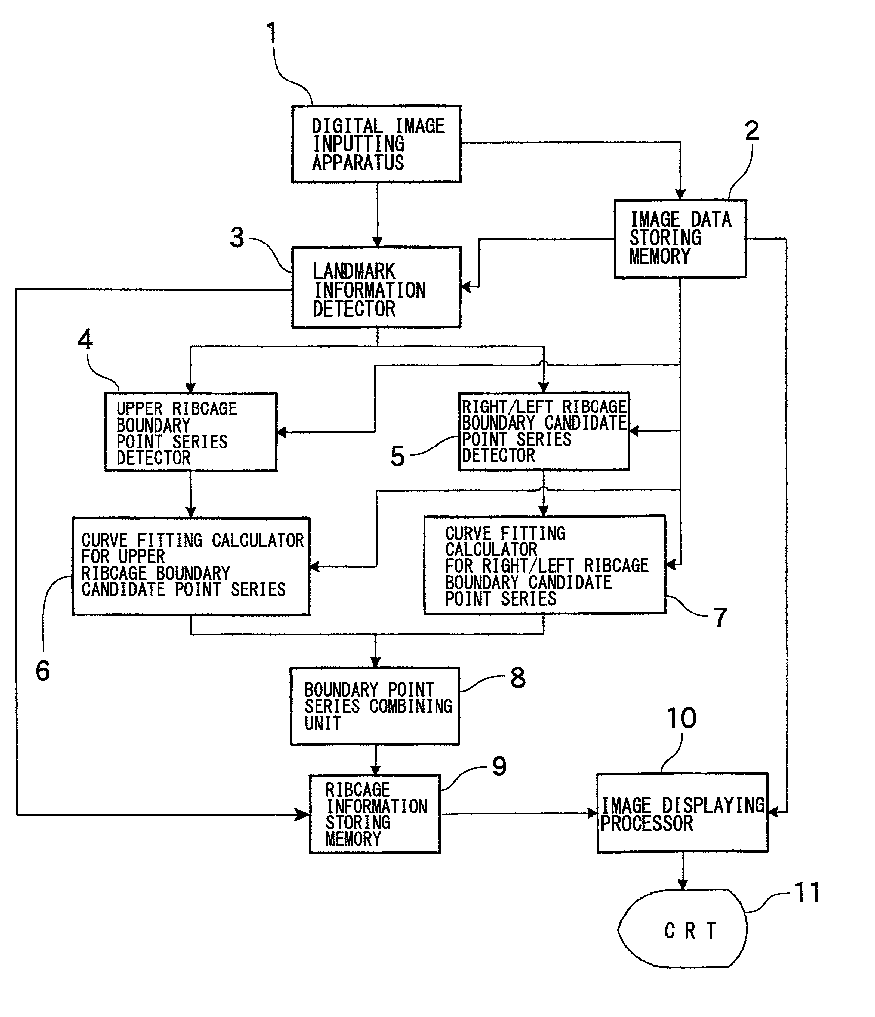
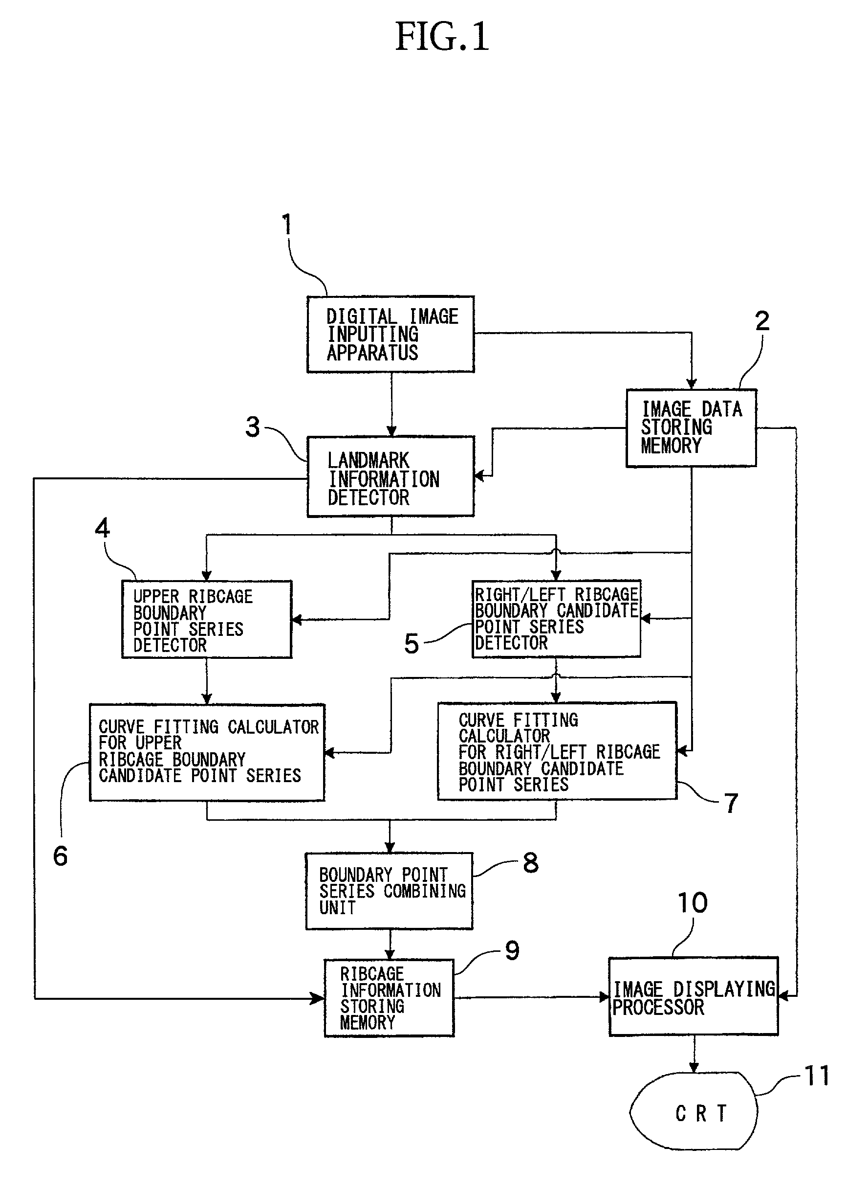
[0039]At first, referring to FIGS. 1 to 4, the configuration of a ribcage boundary detecting system according to an embodiment, which is used for detecting the boundary of a ribcage from a digital chest image, will now be described.

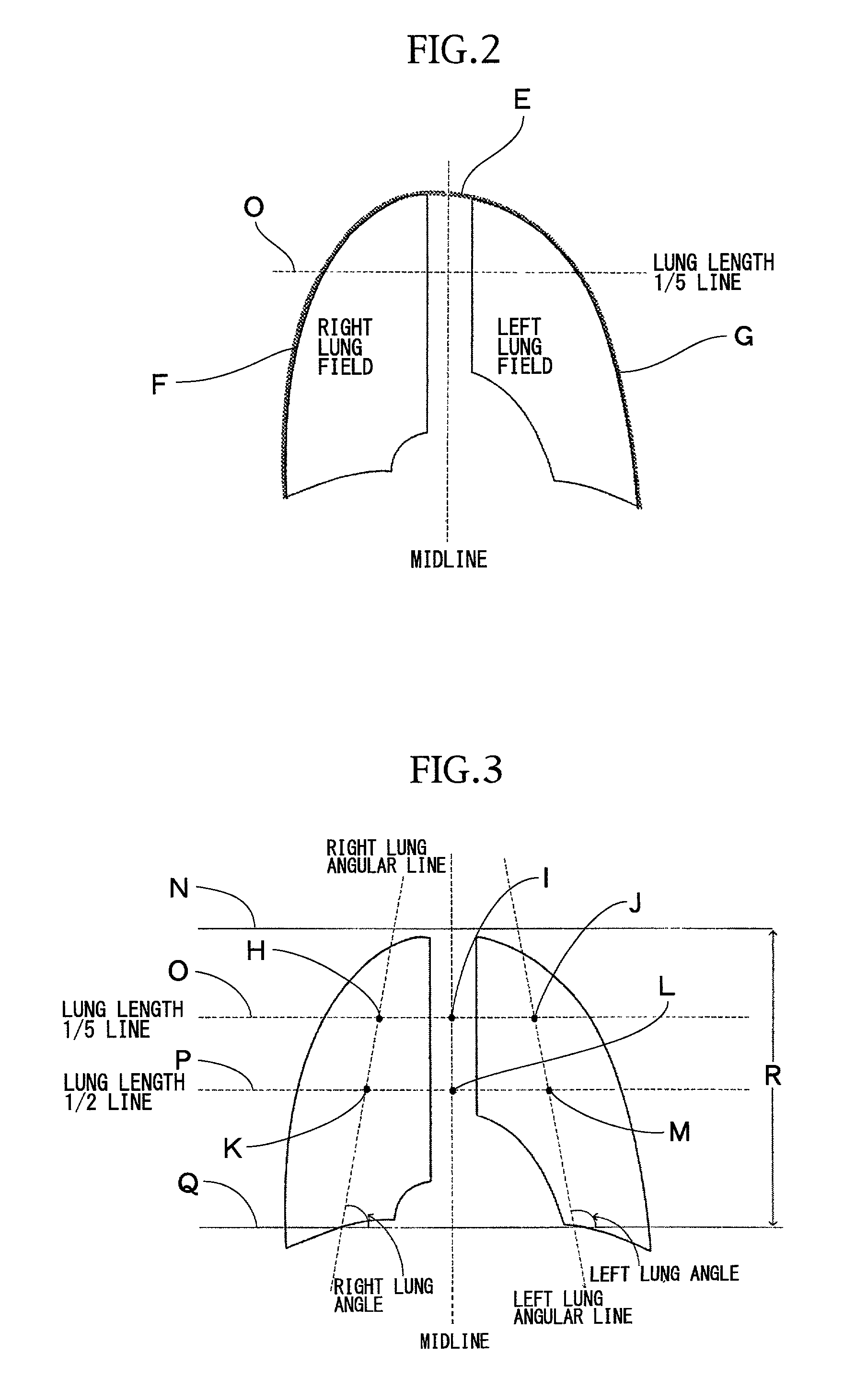
[0040]FIG. 1 shows the entire configuration of the ribcage boundary detecting system according to the present embodiment. FIG. 2 illustrates pieces of ribcage information which are adopted as the anatomical structure information of a chest image. FIG. 3 illustrates pieces of landmark information which composes part of the pieces of the ribcage information. FIG. 4 illustrates lines of boundary candidate points serving as pieces of ribcage boundary information which composes part of the pieces of the ribcage information.

[0041]As shown in these drawings, the ribcage information of a chest image consists of pieces of ribcage boundary informat...


PUM


Abstract

To provide more accurate ribcage boundary information and landmark information, the size of a searching ROI which is mapped on a digital chest image is decided, the ROI is set on the image, an image existing within the region that is set on the image is enhanced, a profile of pixel values existing within the region is obtained, and the obtained profile is analyzed so that candidate points for a ribcage boundary are obtained. The deciding, setting, enhancing, obtaining, and analyzing operations are repeated until a center of each searching ROI advances immediately before a given search limit. As a result, a series of candidate points for the ribcage boundary is provided.

Description

BACKGROUND OF THE INVENTION[0001]1. Field of the Invention[0002]The present invention relates to an image processing technique for automatically and accurately detecting an anatomical configuration from a (PA: postero-anterior) digitized chest radiograph, which is necessary for the computer-aided diagnosis of the chest, and in particular, to an image processing technique that makes it possible to, with higher precision, detect the boundary of a ribcage from a digitized chest radiograph.[0003]2. Description of the Related Art[0004]Digitized chest radiographs have been widely used in the field of computer-aided diagnosis. A wide variety of types of computer-aided diagnosis which is capable of automatically detecting ribcage boundary information and landmark information both specifying anatomical configurations of the chest have been known. One conventional technique is provided by “Xin-Wei Xu and Kunio Doi, Image feature analysis for computer-aided diagnosis: Accurate determination of...

Claims


Application Information

Patent Timeline
Patent Type & Authority Patents(United States)
IPC IPC(8): G06K9/00A61B6/00G06T1/00G06T3/00G06T5/00G06T7/60
CPCG06T7/0083G06T2207/10116Y10S128/922G06T2207/20164G06T2207/30061G06T2207/20104G06T7/12
Inventor OZAKI, OSAMU
Owner MITSUBISHI SPACE SOFTWARE KK
Learn More