Device for processing images, in particular medical images

a technology for processing devices and images, applied in the field of processing devices for images, can solve problems such as difficulties, loss of information about which of the two image series the individual pixels to be represented belong by mixing, and contrast loss due to alpha blending

Active Publication Date: 2006-09-19
SIEMENS HEALTHCARE GMBH
View PDF6 Cites 74 Cited by
  • Summary
  • Abstract
  • Description
  • Claims
  • Application Information

AI Technical Summary

Benefits of technology

[0008]It is therefore an object of the invention to provide an image-processing device which permits meaningful generation of a fusion image, irrespective of the actual gray-value distribution of two images or image series to be fusioned.

Problems solved by technology

On the one hand, the information about which of the two image series the individual pixels to be represented belong to is lost by the mixing.
The contrast loss due to alpha blending is a problem for interpretation of the image data in the fusion image.
Difficulties arise, in particular, whenever images that have been recorded in the same mode are to be fusioned, for example two CT pictures which show the same anatomical area.
This recording of disjoint gray-value ranges, which is a prerequisite for using the threshold mode with a single threshold value, however, is not feasible whenever two images or image series to be fusioned are similar to one another in terms of their gray-value distribution.

Method used


Image

  • Device for processing images, in particular medical images
  • Device for processing images, in particular medical images
  • Device for processing images, in particular medical images

Examples


Embodiment Construction

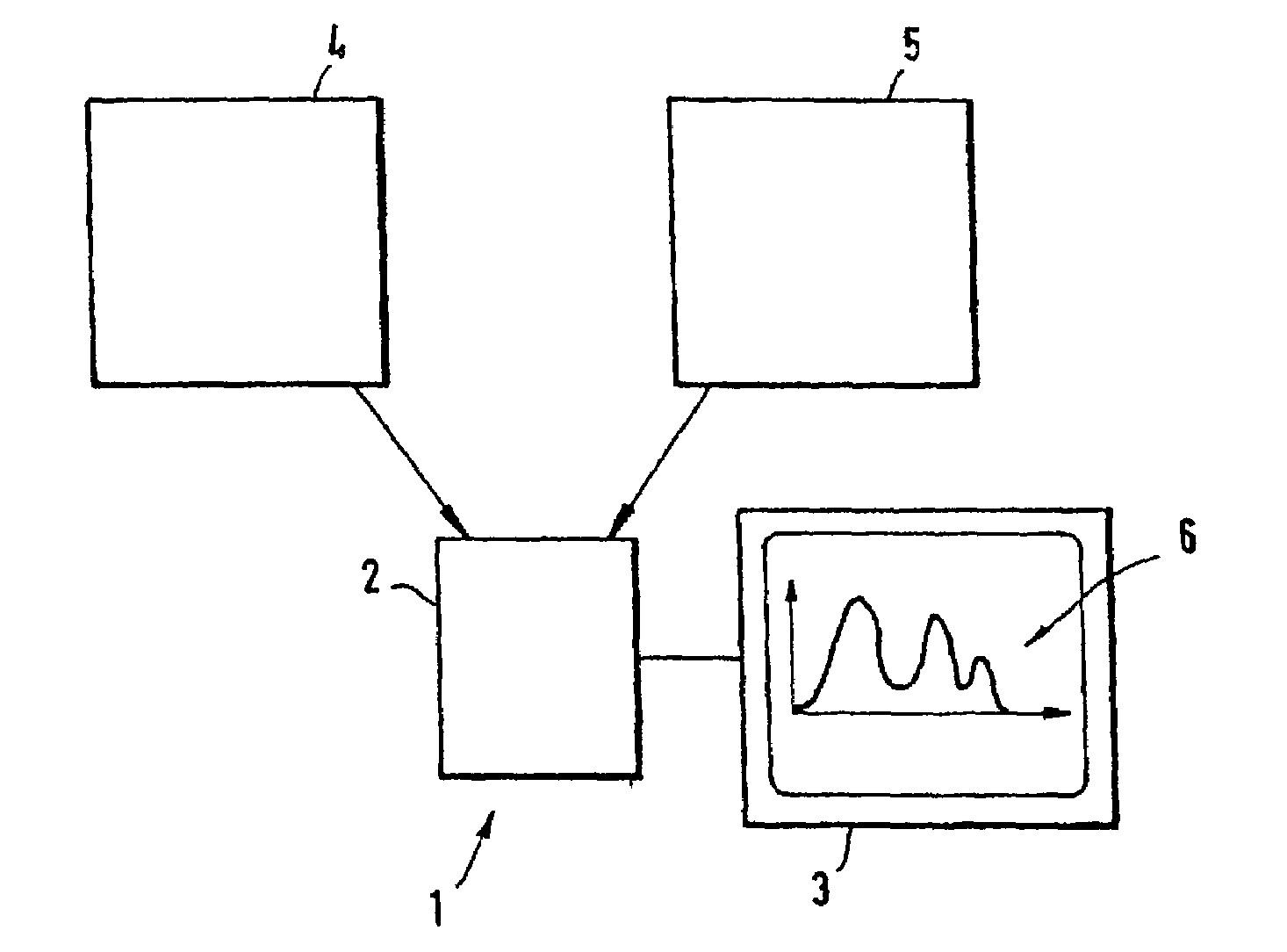
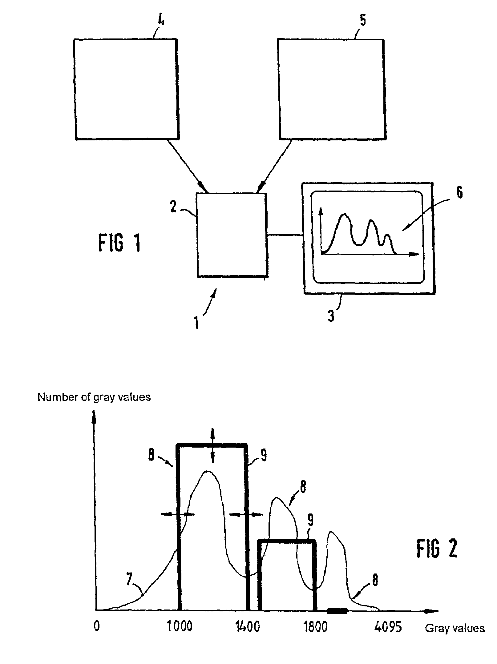
[0032]FIG. 1 shows, in the form of an outline diagram, a device 1 according to the invention for processing images. This device comprises an image-processing computation unit 2 and the monitor 3, on which images and other information can be displayed. In the exemplary embodiment which is shown, the image-processing computation unit 2 contains images which have been recorded using two different recording devices, for example a computer topography instrument 4 and a magnetic resonance instrument 5, and which are to be overlaid. On the monitor 3, it is now possible to represent gray-value histograms 6 for the individual images or image series. The user can modify these gray-value histograms by highlighting markings, so that a meaningful fusion of various images or image series is possible. This will discussed in more detail below. The fusion of the images is carried out by the image-processing computation unit, the fusion result being subsequently output in turn on the monitor 3. Of co...


PUM


Abstract

A device for generating fusion images by fusioning two images, in particular medical images, comprising an image-processing computation unit (2) for fusioning the images, as well as a monitor (3) connected thereto for image output, a gray-value histogram (7, I, II) being displayable on the monitor (3) for each image of the images to be fusioned, which device is designed in such a way that, in each gray-value histogram (7, I, II) of the images to be fusioned, one or more gray-value ranges can be selected by user-controlled highlighting of one or more markings (8) on the monitor (3), as well as for generating the fusion image with the aid of the selected gray-value ranges.

Description

FIELD OF THE INVENTION[0001]The invention relates to a device for processing images, in particular medical images, a gray-value histogram being displayable on a monitor for an image.BACKGROUND OF THE INVENTION[0002]Especially in the field of medicine, image fusion methods are being used for better diagnosis of a possible pathological state. The purpose of image fusion is to combine and visually overlay medical images or image series of a patient, which contain supplementary information to one another. The method most commonly used at present for visually overlaying image series or images is so-called alpha blending. In this case, the gray values of the two images or image series are weighted with a blending factor (alpha value) and the weighted average value of the two images to be fusioned is represented for each image point. By varying the alpha value, it is possible to vary the relative weighting of the images or image series in relation to one another. Discrimination of the two ...

Claims


Application Information

Patent Timeline
Patent Type & Authority Patents(United States)
IPC IPC(8): G06K9/36G06T3/00G06T5/40G06T15/10G06T15/50H04N1/387
CPCG06T5/40G06T5/50G06T15/10G06T11/00G06T15/503G06T2207/20221G06T2207/30004
Inventor DITT, HENDRIKRAHN, NORBERTWACH, SIEGFRIED
Owner SIEMENS HEALTHCARE GMBH
Why Eureka
Try Eureka