Radiation image recording device

a technology of imaging device and recording device, which is applied in the direction of material analysis using wave/particle radiation, instruments, applications, etc., can solve the problems of complex process and complex process

Inactive Publication Date: 2006-11-28
SIEMENS HEALTHCARE GMBH
View PDF12 Cites 41 Cited by
  • Summary
  • Abstract
  • Description
  • Claims
  • Application Information

AI Technical Summary

Benefits of technology

[0019]The retaining devices can thereby be configured as handles, the height of which can be varied, so that people of differing heights can be secured optimally. It is also possible to design the retaining means as corresponding straps, etc., which are used to belt the patient firmly in position.
[0020]A radiation-transparent plate is also expediently provided on the platform on the side facing the radiation receiver, to prevent the patient coming into contact with the radiation receiver.

Problems solved by technology

This process is elaborate and complex and the storage plates require subsequent development, which takes a relatively long time, so diagnosis can not take place at the same time as recording.

Method used


Image

  • Radiation image recording device
  • Radiation image recording device
  • Radiation image recording device

Examples


Embodiment Construction

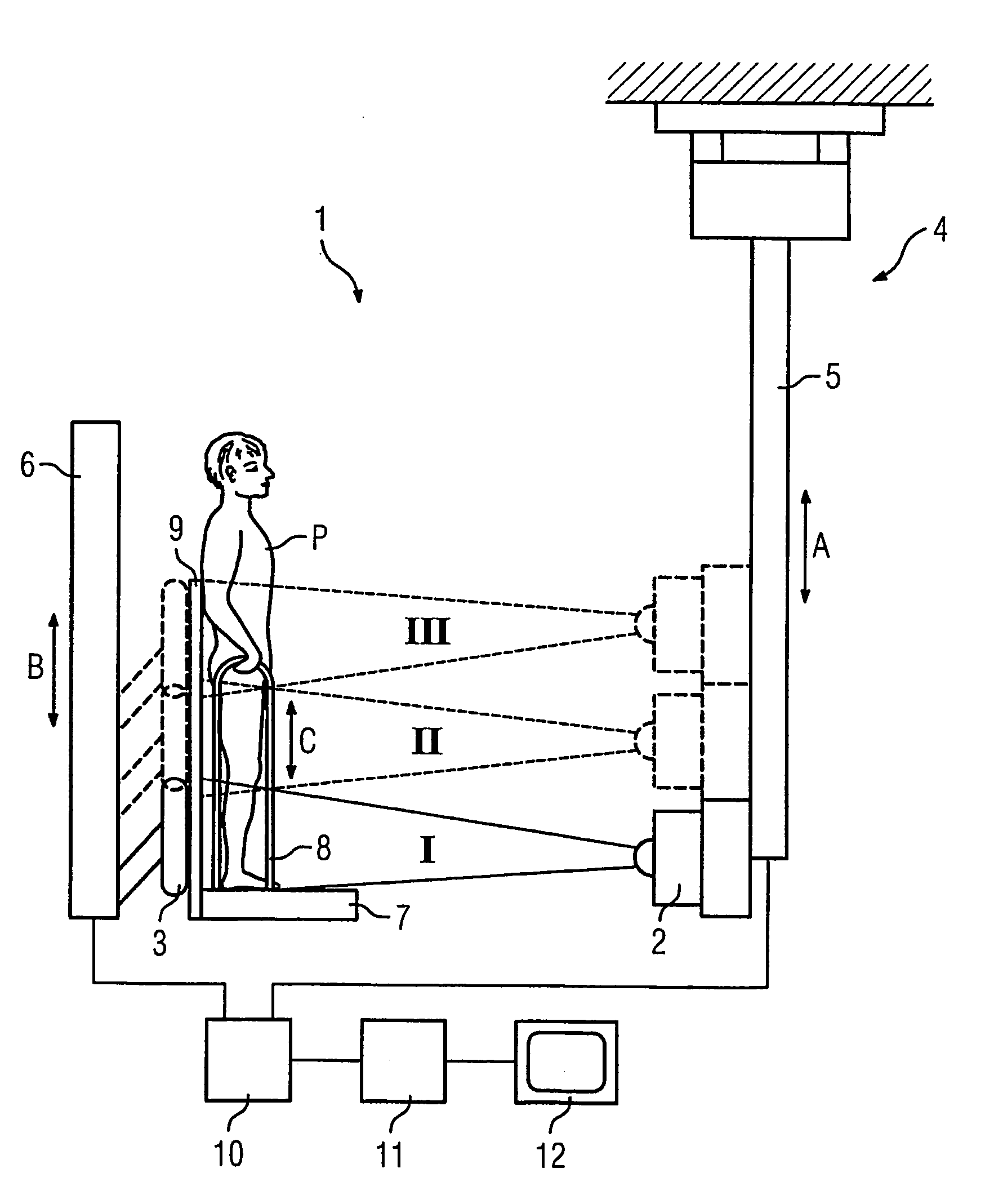
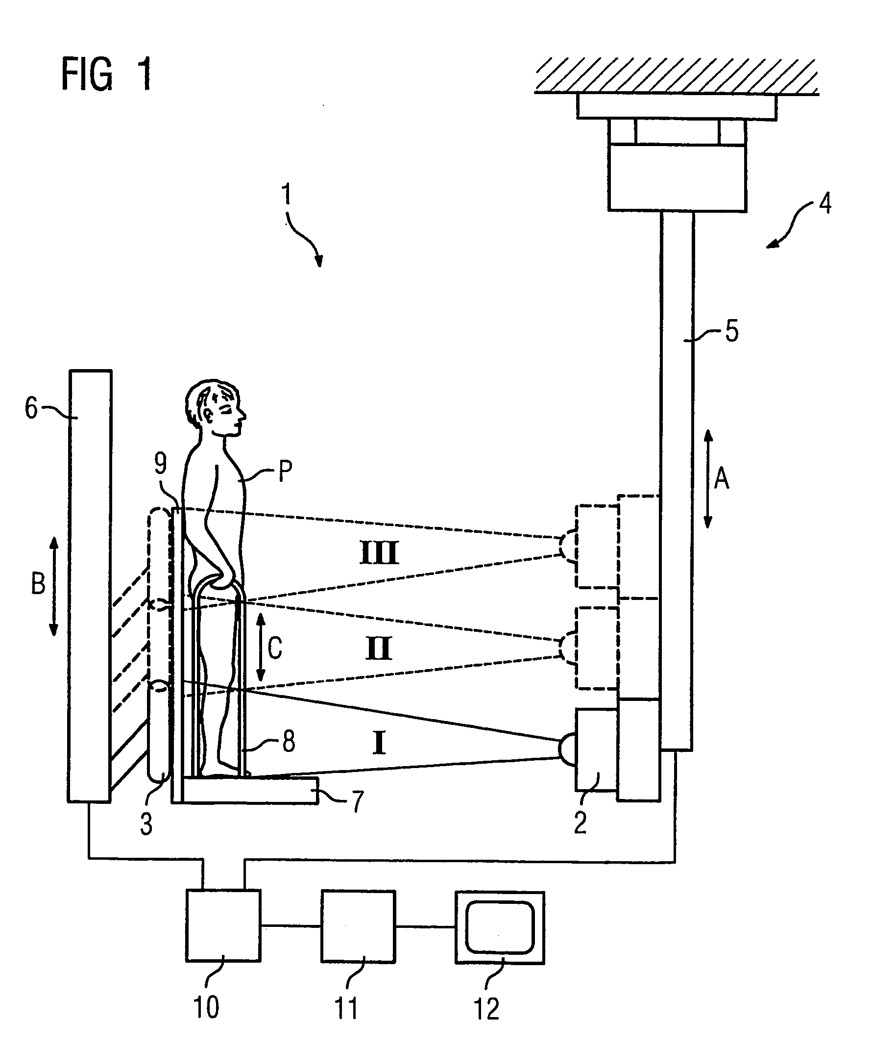
[0024]FIG. 1 shows a radiation imaging device 1 according to the invention, comprising a radiation source 2, in this case an X-ray emitter, and a radiation receiver 3, in this case a digital solid state detector. The radiation source 2 is arranged on a gantry 4 with a telescopic bar 5 and can therefore be moved vertically as shown by the double arrow A. The same applies to the radiation receiver 3. This is also arranged on a gantry 6 and can also be moved vertically, as shown by the double arrow B. While the gantry 4 is supported on the ceiling, the gantry 6 is a floor gantry.

[0025]A platform 7 is provided in the vicinity of the radiation receiver, on which the patient P has to stand for the recording. Retaining means 8 in the form of vertically movable handles (see double arrow C) are arranged on both sides of the platform 7, which the patient can hold on to, as said patient has to stand still during imaging. A radiation-transparent plate 9 is also provided at the back, arranged th...


PUM

PropertyMeasurementUnit
heightaaaaaaaaaa
surface areaaaaaaaaaaa
areaaaaaaaaaaa

Abstract

Disclosed is a radiation image recording device comprising a source of radiation and a radiation receiver which are movable in a vertical direction in order to be positioned relative to a standing patient, and an image processing device for creating an output image based on the recorded image data. The source of radiation and the radiation receiver are movable in a controlled manner into successive image recording positions via a control device so as to record an area of analysis which exceeds the height of the active area of the digital radiation receiver, one radiation image being recorded in each image recording position. The positions are defined in such a way that the recorded radiation images cover the area of analysis while the image processing device is embodied so as to create a full image representing the entire area of analysis based on the image data of the individual radiation images.

Description

CROSS REFERENCE TO RELATED APPLICATIONS[0001]This application claims priority to the German application No. 10244609.1, filed Sep. 25, 2002 and to the International Application No. PCT / DE03 / 03138, filed Sep. 22, 2003 which are incorporated by reference herein in their entirety.FIELD OF INVENTION[0002]The invention relates to a radiation imaging device with a radiation source and a radiation receiver, which can be moved in a vertical direction to be positioned in relation to a standing patient, and with an image processing device for producing an image that can be output based on the recorded image data.BACKGROUND OF INVENTION[0003]In modern X-ray-based diagnostics it is increasingly frequently necessary to examine large examination areas, e.g. the entire spinal column or the leg area to diagnose bone positions. The patient is hereby scanned in a standing position with the radiation imaging device, i.e. a conventional X-ray device comprising an X-ray tube and an X-ray radiation recei...

Claims


Application Information

Patent Timeline
Patent Type & Authority Patents(United States)
IPC IPC(8): G01N23/06A61B6/00A61B6/02
CPCA61B6/4225A61B6/4464A61B6/5241
Inventor ATZINGER, FRANZHERRMANN, ROLANDKOHLER, VOLKMARSCHRAMM, HELMUTH
Owner SIEMENS HEALTHCARE GMBH
Why Eureka
Try Eureka