Methods and apparatus for accessing and stabilizing an area of the heart

a technology of accessing and stabilizing an area of the heart, applied in the direction of internal electrodes, therapy, surgery, etc., can solve the problems of not providing access, unable to reliably maintain a vacuum seal against the pericardium when such treatment devices are advanced, and unable to provide access

Active Publication Date: 2006-12-05
MEDTRONIC INC
View PDF33 Cites 243 Cited by
  • Summary
  • Abstract
  • Description
  • Claims
  • Application Information

AI Technical Summary

Benefits of technology

[0020]The present invention is preferably embodied in an epicardial suction tool for engaging the epicardium having an elongated tool body extending between a tool body proximal and distal ends and encloses a tool body lumen. At least one suction pad supported by a suction pad strut extends distally to a distal end surface of the tool body distal end. Suction is applied through suction lumens to suction ports of the suction pad that fix to the epicardium so that the suction tool distal end tracks movement of the epicardium. Various procedures can be performed and medical devices, instruments, drugs can be advanced through the tool body lumen without interrupting suction.

Problems solved by technology

Although theoretically plausible, the ability to reliably maintain a vacuum seal against the pericardium when such treatment devices are advanced can be problematic.
Epicardial pacing and cardioversion / defibrillation leads were the first to be implanted widely, because endocardial leads lacked effective active or passive fixation mechanisms and relied upon relatively stiff lead bodies that cause perforations and dislodgement of the distal electrode(s) or fracture of the lead conductor.
These procedures involve major surgery that may be painful and dangerous for the patient and can be costly.
The subxiphoid approach, moreover, only permits limited access to the anterolateral surface of the left ventricle and does not provide any access at other locations of the left ventricle or the left atrium.
Endocardial pacing electrodes cannot be implanted directly within the left heart chambers due to risk of thrombus formation about the lead body and electrode surfaces.
Implanted objects often cause minor blood clots and thrombus to form in the blood.
Any blood clots or thrombi, however minor, that form in the left atrium and ventricle could have serious consequences if they were to break free and be swept into the brain and cause a stroke.
At times it is difficult to advance the pacing and / or cardioversion / defibrillation electrodes into or to a desired site of the coronary sinus or great vein.
And, the lead bodies tend to fibrose into the coronary sinus or great vein over time and become difficult or impossible to remove if it becomes desirable to do so.

Method used


Image

  • Methods and apparatus for accessing and stabilizing an area of the heart
  • Methods and apparatus for accessing and stabilizing an area of the heart
  • Methods and apparatus for accessing and stabilizing an area of the heart

Examples


Embodiment Construction

[0038]In the following detailed description, references are made to illustrative embodiments for carrying out the invention. It is understood that other embodiments may be utilized without departing from the scope of the invention.

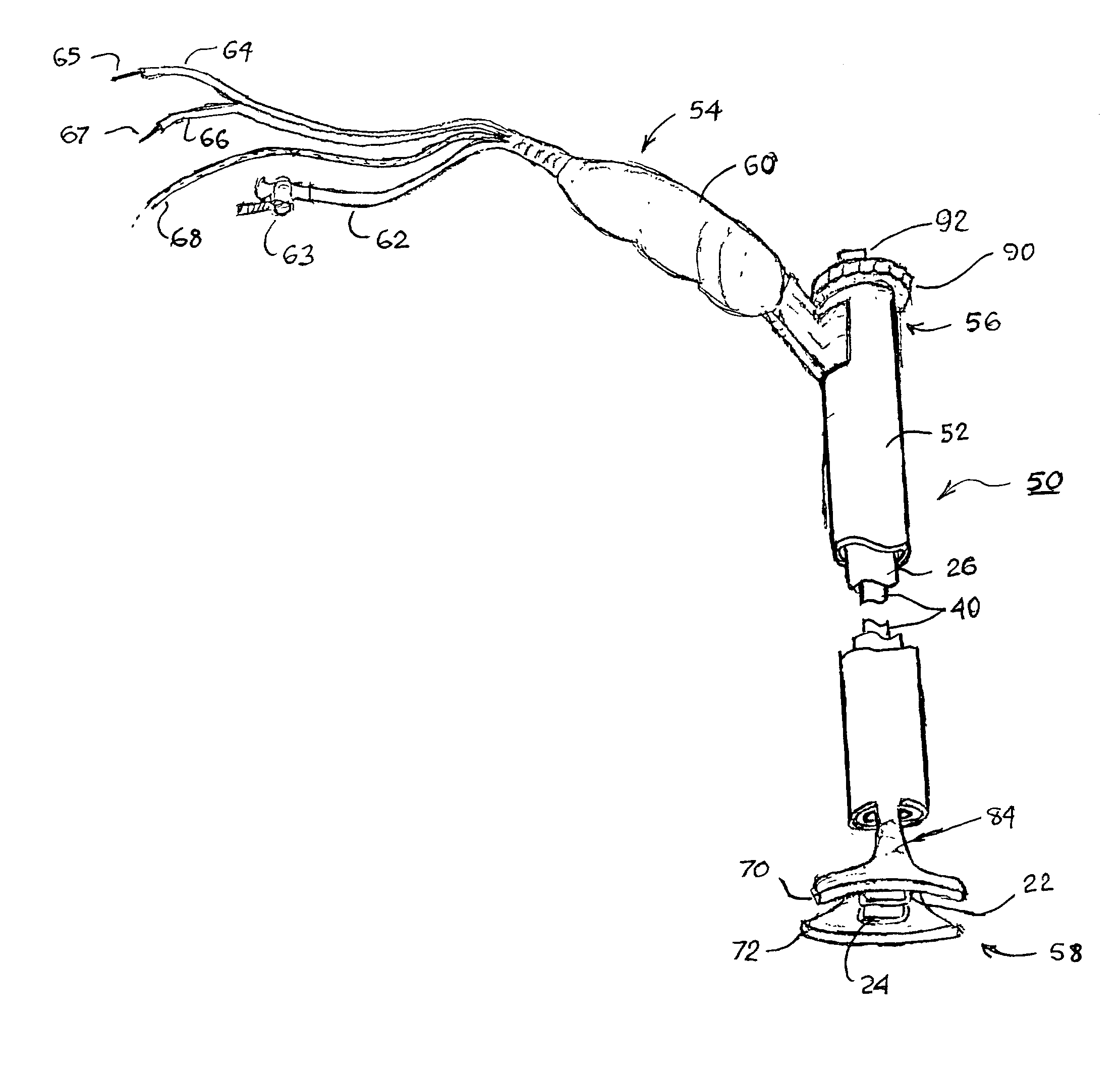
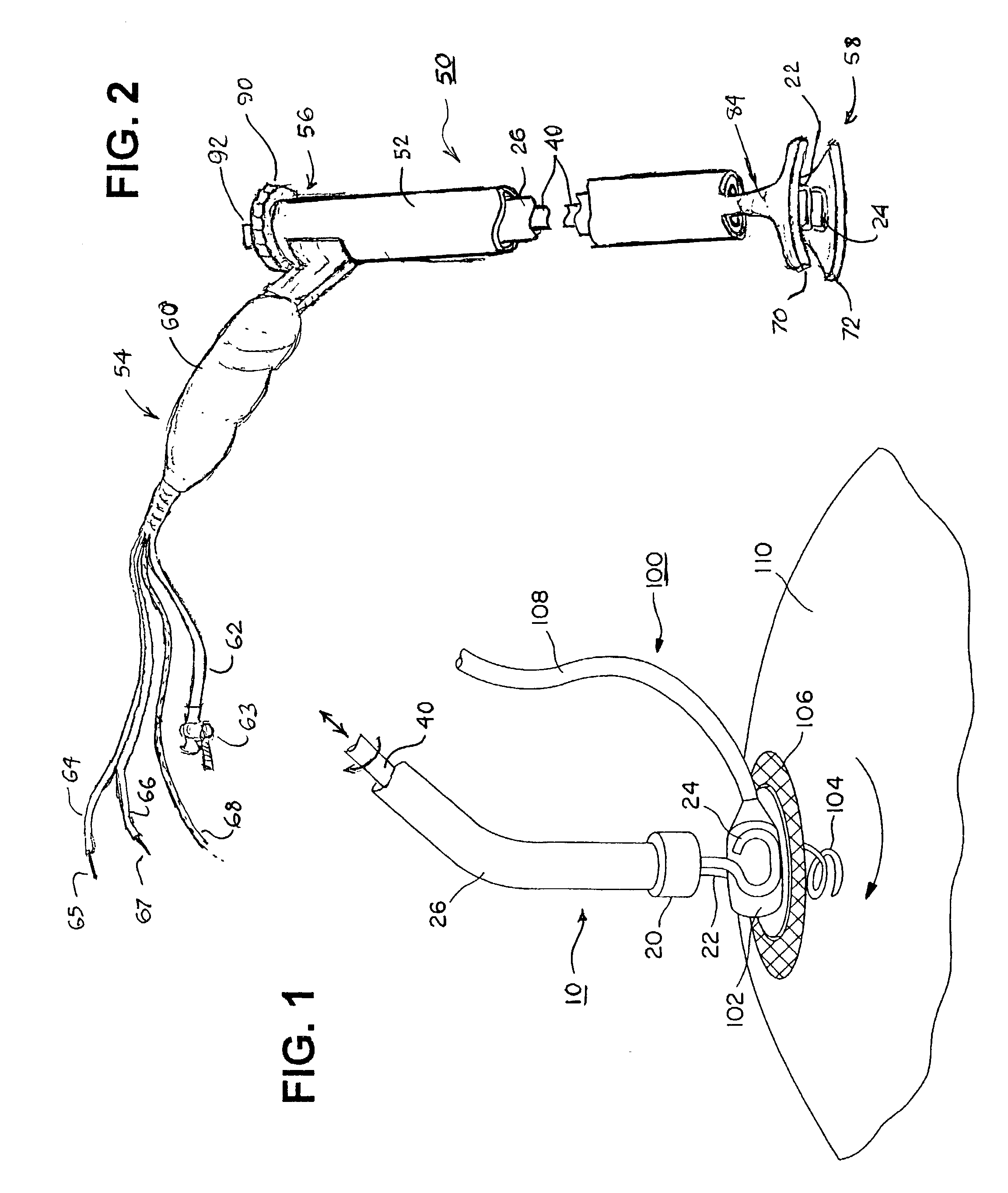
[0039]FIG. 1 is a plan view of an implantation tool 10 of the type disclosed in the above-referenced '526 patent, which is incorporated into a preferred embodiment of the epicardial lead installation tool of the present invention, gripping the electrode head 102 of an epicardial screw-in lead 100 to screw the helical electrode 104 through the epicardium accessed through a surgical incision into the myocardium 110. The implantation tool 10 comprises outer shaft tubing 26 having a lumen extending between the device proximal end (not shown) and the distal nose cap 20. An internal shaft or cable 40 extends through the lumen of the outer shaft tubing 26 between a pair of spaced apart tongs 22 and 24 and a proximal handle assembly (not shown) that enables select...


PUM


Abstract

An epicardial lead installation tool having an elongated tool body extending between a tool body proximal and distal ends and encloses a tool body lumen. At least one suction pad supported by a suction pad strut extends distally to a distal end surface of the tool body distal end. A lead implantation tool extending between implantation tool proximal and distal ends is inserted through the tool body lumen to dispose the implantation tool distal end proximate to the tool body distal end. The lead implantation tool distal end is adapted to engage the distal electrode head of an epicardial lead to enable the extension of the assembly of the of the lead installation tool, the lead implantation tool and the epicardial lead through a skin incision and to apply the suction pad against the epicardium. Suction is applied through the suction pad to affix it to the epicardium while the implantation tool proximal end is manipulated to affix the distal fixation mechanism to the myocardium at the implantation site.

Description

FIELD OF THE INVENTION[0001]The present invention pertains methods and apparatus for accessing and stabilizing an area of the heart, particular a portion of the heart wall to enable performance of a surgical procedure through minimally invasive surgical techniques.BACKGROUND OF THE INVENTION[0002]Many minimally invasive surgical techniques and devices designed to reduce morbidity, expense, and the trauma associated with open-chest cardiac surgery have revolutionized cardiac surgery. Recent advances in endoscopic and thoracoscopic instruments and percutaneous access to a patient's thoracic cavity have made minimally invasive surgery possible. The advent of thoracoscopy in cardiac surgery has shown promise as a technique to enable surgeons to implant epicardial leads without sternotomy or thoracotomy. Thoracoscopy normally involves making small incisions between the ribs into the chest cavity and passing tubular ports Through the incisions. Illumination devices, cutting instruments, s...

Claims


Application Information

Patent Timeline
Patent Type & Authority Patents(United States)
IPC IPC(8): A61B1/00A61N1/05
CPCA61N1/0587
Inventor GUENST, GARY W.FOREST, GERARD C.
Owner MEDTRONIC INC
Why Eureka
Try Eureka