Method of analyzing cell samples, by creating and analyzing a resultant image

a cell sample and resultant image technology, applied in the field of cell sample analysis, can solve the problems of mismatching of spots, inaccuracy of further analyses, and time-consuming above-described analysis steps in the method known from wo-96/33406, and achieve the effect of improving accuracy

Inactive Publication Date: 2006-12-26
GE HEALTHCARE BIO SCI CORP
View PDF5 Cites 78 Cited by
  • Summary
  • Abstract
  • Description
  • Claims
  • Application Information

AI Technical Summary

Benefits of technology

[0018]Another object of the present invention is to achieve a higher degree of accuracy in the anal

Problems solved by technology

The above described analysis step in the method known from the WO-96/33406 is sometimes very time-consuming.
One drawback is the risks that spots are mismatched, i.e. spots believed to be corresponding in the two images are not.
Another drawback might be that the further anal

Method used


Image

  • Method of analyzing cell samples, by creating and analyzing a resultant image
  • Method of analyzing cell samples, by creating and analyzing a resultant image
  • Method of analyzing cell samples, by creating and analyzing a resultant image

Examples


Embodiment Construction

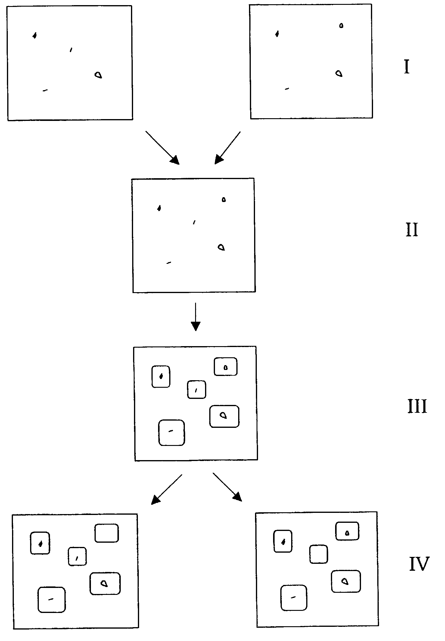
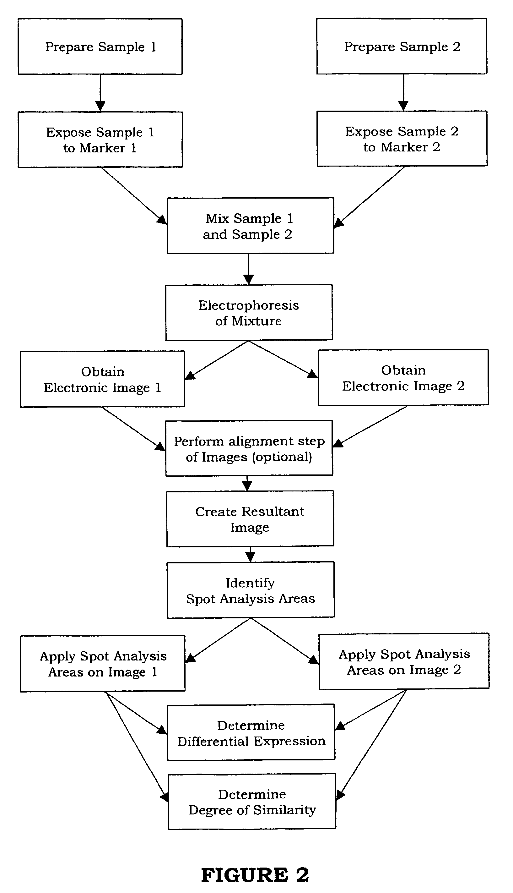
[0037]FIG. 2 shows flow diagram illustrating the method according to the invention. The different steps illustrated in the flow diagram are described below with references to all the figures.

[0038]Throughout the description, in the example illustrating the invention is the separation step preferably performed by electrophoresis. However, the invention is equally applicable in any other separation method.

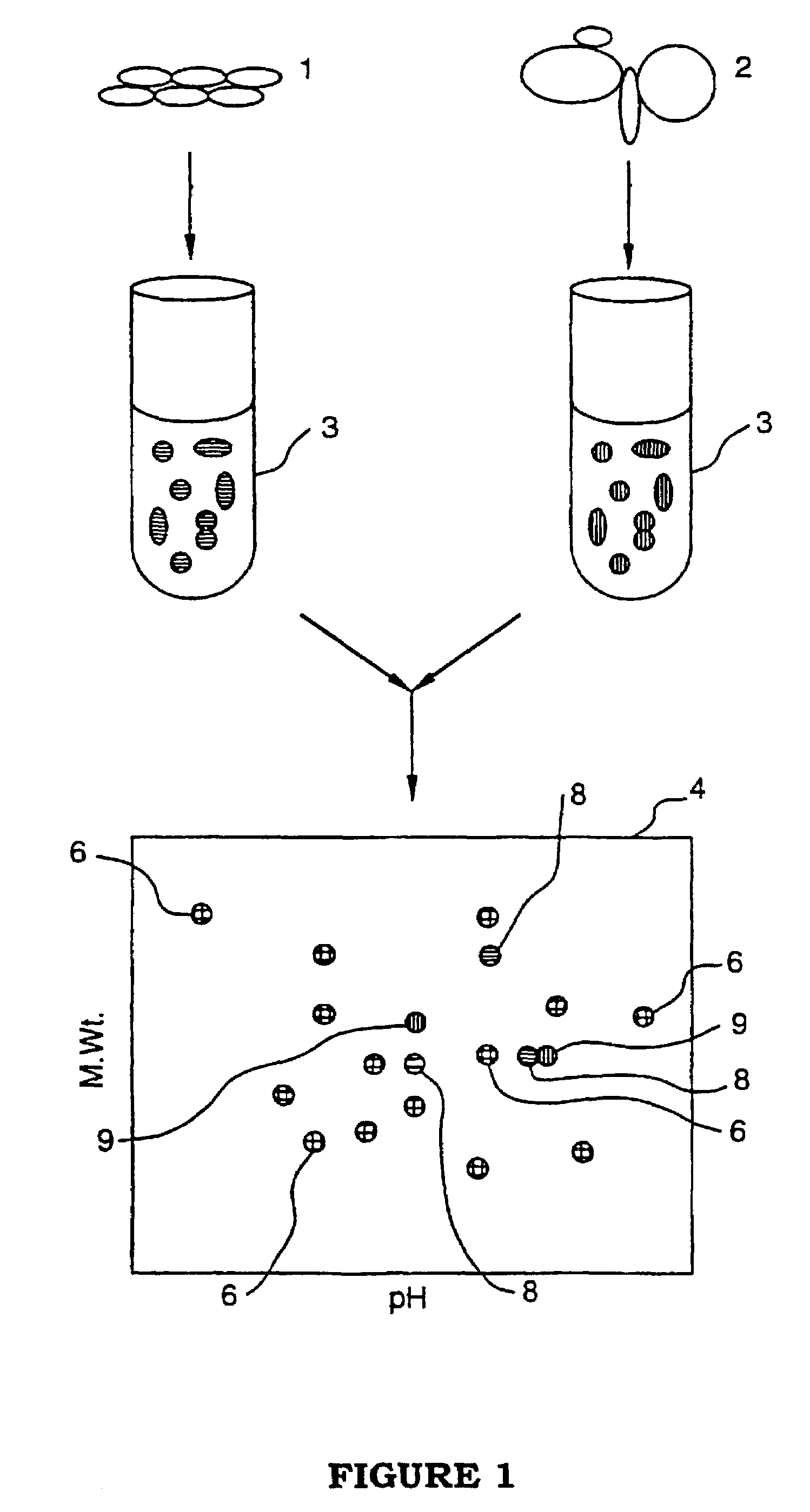
Preparation and Mixing Step

[0039]According to a preferred embodiment of the invention the process of the present invention employs a matched set of dyes wherein each dye in the set is generally equal to the other dyes in ionic and pH characteristics, and chemical reactivity for covalent attachment to proteins, yet fluoresces at a different wavelength, thereby exhibiting a different color luminescence when viewed. The dyes are preferably roughly equal in molecular weight, but need not be. Each one of the dyes within the matched set of dyes is used to label proteins in a different one ...


PUM

PropertyMeasurementUnit
Areaaaaaaaaaaa
Stretching forceaaaaaaaaaa
Luminescenceaaaaaaaaaa

Abstract

A method is provided for comparing multiple samples of cell extract containing a plurality of components. The method comprises the steps of preparing at least two samples of cell extract from at least two groups of cells and of exposing each of said sample of said cell extract to a different one of a set of matched markers, e.g. luminescent markers, to bind the marker to the cell extract to label the cell extract, each marker within said set of markers being capable of binding to the cell extract and can be individually detected from all other markers within said set. The samples are then mixed to form a mixture and said mixture is electrophoresed to separate the components within the cell extract. At least two electronic images of the electrophoresed mixture are obtained (I) by detection of the individual markers, each image being represented by detection of a marker different from the others. One resultant electronic image (Ires) of the obtained at least two electronic images is created (II) and analyzed in order to identify spot analysis areas (III). The identified spot analysis areas are applied on the respective at least two electronic images for evaluating said areas in order to detect spots representing components of said cell extracts (IV).

Description

BACKGROUND OF THE INVENTION[0001]1. Field of the Invention[0002]The present invention relates to a method of comparing multiple samples of cell extracts for detecting differences in the protein composition of cells and cells extracts, and more particularly, to a method utilizing matched pair of labeling reagents for detecting such differences. By the expression samples of cell extracts is meant any sample that can be subject to the analysis method outlined in the present application.[0003]2. Background of the Invention[0004]Researchers studying various aspects of cell biology use a variety of tools to detect and monitor differences in cell structure, function and development. An essential part of studying cells is studying the differences and similarities in the protein composition between the different cell types, stages of development and condition. Determining differences in the protein content between normal and cancerous cells or wild type and mutant cells, for example, can be ...

Claims


Application Information

Patent Timeline
IPC IPC(8): G06K9/00G01N33/48G01N27/447G06T7/00
CPCG06K9/00127G06T7/0012G06V20/69
Inventor SLOGE, ERIKSODERMAN, TOBIASBJORKESTEN, LENNART
Owner GE HEALTHCARE BIO SCI CORP
Why Eureka
Try Eureka