
Medical tomography apparatus for generating a 2D image from a 3D dataset of a tomographic data

a tomography apparatus and 3d dataset technology, applied in tomography, applications, instruments, etc., can solve the problems of multi-planar reconstruction, difficult to define or determine a curved evaluation surface in a number of dimensions, and the presentation of the original tomogram often has little diagnostic utility or does not allow certain viewing modes

Inactive Publication Date: 2007-03-20
SIEMENS AG
View PDF14 Cites 36 Cited by
  • Summary
  • Abstract
  • Description
  • Claims
  • Application Information

AI Technical Summary

Benefits of technology

[0017]It is an advantage of the invention that radiological questions as derived from presentations in pathological textbooks and reference books can be directly replicated. Such books often show curved surfaces in a plane projection in order to make the relevant information optimally accessible to the eye of the observer. Structures of the patient to which pre-defined surfaces can be adapted are, for example, the spinal column, structures of the knee joint, the course of specific blood vessels (carotid artery, aorta), the course of specific nerves (spinal channel), etc.
[0018]In particular, a surface curved in multiple dimensions is selected as predefined surface. The surface thus can have an arbitrary curvature.
[0019]In a preferred embodiment, the evaluation surface is defined by scaling the pre-defined surface with a measured quantity of the patient. As a result thereof, it becomes possible to store the pre-defined surfaces as quasi-standardized surfaces in the library that merely reproduce the shape of the evaluation surface. Only a comparatively small number of surfaces thus need to be stored. The measured quantity of the patient that is employed can either be separately measured or can be a measured quantity that can likewise be obtained by measurement using the tomography apparatus, for example the head diameter of the patient.
[0020]In an embodiment of the method, a digital marking attached to the predefined surface and that corresponds to a characteristic structure feature of the patient is aligned at the structure feature visible in the 3D dataset for the correct positioning of the evaluation surface. The alignment occurs either interactively by the operator or automatically via an image analysis process, for example with “mutual information”.
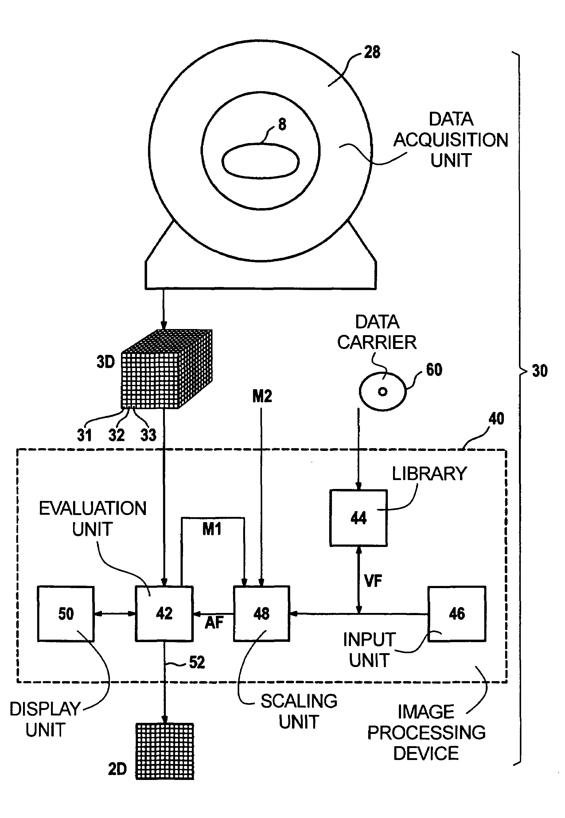
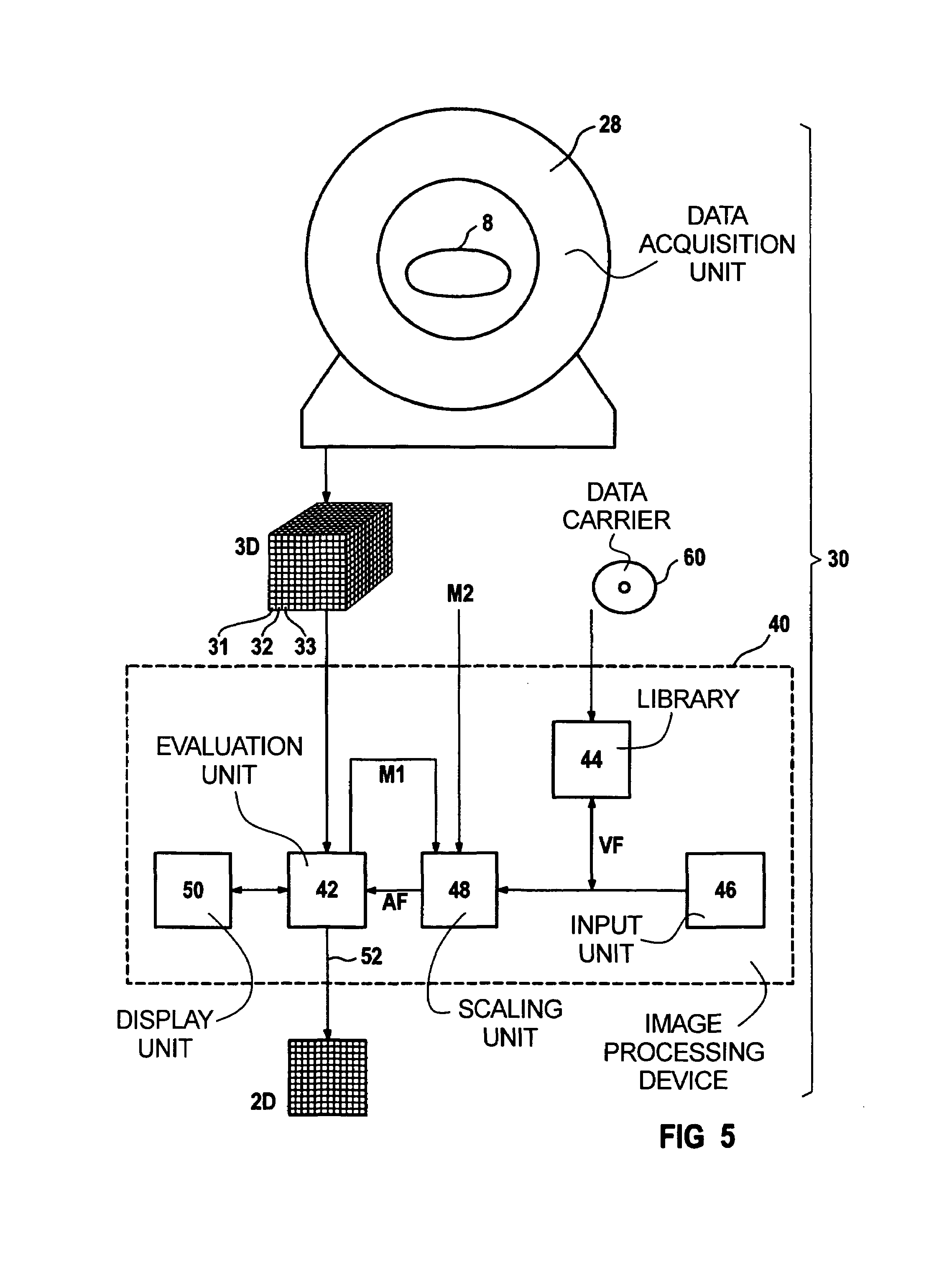
[0021]The above object also is achieved in accordance with the invention medical tomography apparatus with a library having predefined surfaces for defining the shape of the evaluation surface, with one of the predefined surfaces being selectable via an input unit.
[0022]The advantages, embodiments and developments described above for the method apply analogously to the tomography apparatus of the invention.

Problems solved by technology

The presentation of the original tomograms often has little diagnostic utility or do not allow certain viewing modes at all.
Multi-planar reconstruction is particularly tedious for medical personnel when a secondary section, i.e. an evaluation surface proceeding through the grid, must be defined with high complexity.
For example, it is tedious to define or determine a curved evaluation surface in a number of dimensions.
Methods that require an input of a parameter (for example, density value) of a specific surface are susceptible to error and / or require experience, or trial and error.

Method used


Image

  • Medical tomography apparatus for generating a 2D image from a 3D dataset of a tomographic data
  • Medical tomography apparatus for generating a 2D image from a 3D dataset of a tomographic data
  • Medical tomography apparatus for generating a 2D image from a 3D dataset of a tomographic data

Examples


Embodiment Construction

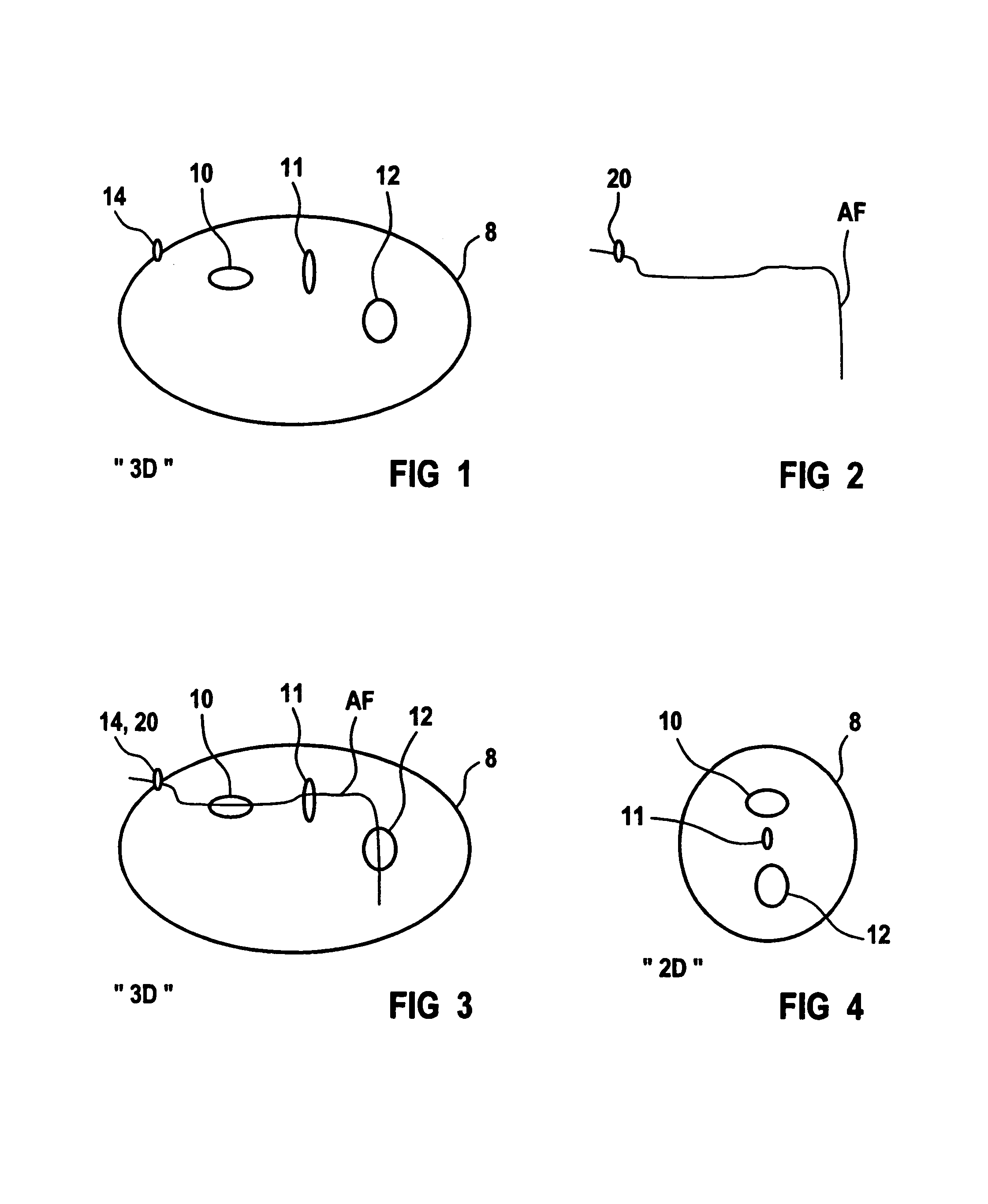
[0036]FIG. 1 shows a real anatomy of a patient 8 acquired in a 3D dataset 3D with structures 10, 11, 12 shown as examples, such as nerves, blood vessels, bones, etc. An emphasized, characteristic or easily identifiable structure feature 14 is likewise shown.

[0037]It is clear that the 3D dataset 3D has been shown only two-dimensionally in FIG. 1 (as well as in FIG. 3).

[0038]FIG. 2 indicates an evaluation surface AF that was acquired from a previously generated, predefined surface VF (see FIG. 5) that was stored in a library 44 (see FIG. 5). Just like the predefined surface VF, the evaluation surface AF contains a digital marking 20 that corresponds to the characteristic structure feature 14, for example a bone part that can be especially easily recognized.

[0039]The logical anatomical landmark or the digital marking 20 is brought into coincidence in FIG. 3 with the anatomical landmark or the structure feature 14 of the patient 8. For correct alignment and positioning of the evaluation...


PUM


Abstract

A medical tomography apparatus and method, a 3D dataset is generated in as picture elements arranged in a three-dimensional grid during the examination of a patient. Predefined surfaces are stored in a library and, dependent on the desired examination, one of the predefined surfaces can be selected with an input unit and thus the shape of an evaluation surface can be defined. Picture elements of the 3D dataset along the evaluation surface and used for constructing a two-dimensional image. A pre-defined surface is selected from the library and is employed for determining the shape of the evaluation surface. The method and the apparatus can be particularly employed in a simple way for the multi-planar reconstruction of a two-dimensional image along a multiply or arbitrarily curved surface.

Description

BACKGROUND OF THE INVENTION[0001]1. Field of the Invention[0002]The present invention is directed to a method for generating a two-dimensional image from a 3D dataset of a tomography apparatus for the medical examination of a patient, wherein the 3D dataset contains picture elements arranged is three-dimensional grid, and wherein an evaluation surface proceeding through the grid is defined and the picture elements along the evaluation surface are employed for constructing the two-dimensional image. The invention is also directed to a medical tomography apparatus with which a 3D dataset is generated in as picture elements in a three-dimensional grid during the examination of a patient, having an input unit for defining an evaluation surface proceeding through the grid, and having an evaluation unit for constructing a two-dimensional image from the 3D dataset using the picture elements along the evaluation surface. The invention is also directed to a data carrier for such a method.[00...

Claims


Application Information

Patent Timeline
Patent Type & Authority Patents(United States)
IPC IPC(8): G06K9/00A61B6/00G06K9/36G01R33/32A61B5/00A61B5/055A61B6/03A61B8/00G01T1/161G01T1/17G06T11/00G06T15/08G06T19/00
CPCG06T15/08G06T19/00A61B6/506G06T2210/41G06T2219/008
Inventor FABER, ROLANDKUTH, RAINER
Owner SIEMENS AG
Learn More