Neuromuscular monitoring using phonomyography

a technology of phonomyography and neutrus, applied in the field of phonomyography, can solve the problems of reducing the knowledge about the action of muscle relaxants, putting patients at great risk of respiratory complications, and reducing the effectiveness of surgery,

Inactive Publication Date: 2007-06-26
UNIV DE MONTREAL +2
View PDF2 Cites 89 Cited by
  • Summary
  • Abstract
  • Description
  • Claims
  • Application Information

AI Technical Summary

Problems solved by technology

A patient who is extubated when still partially relaxed is at great risk of respiratory complications.
Also, a patient incompletely relaxed during surgery can endanger the success of surgery.
Unfortunately, despite these facts, knowledge about the action of muscle relaxants is still quite limited and the tools to measure their function in daily routine are even more limited.
This discovery meant that the monitoring of only one, easily accessible muscle, such as the adductor pollicis muscle, was no longer valid to reflect muscle relaxant action in the human body. p The last 15 years were spent to develop methods for monitoring different muscles, such as the larynx, the diaphragm or the eye muscles.
Although these discoveries were important for research, clinical monitoring of more central, but nevertheless important muscles during surgery (e.g. abdominal surgery) and / or anesthetic relaxation (e.g. intubation) was impaired by the fact that most methods were unsuited for clinical use.
A fundamental problem of all these research efforts of comparing neuromuscular blockade at different muscles remained that the gold standard of neuromuscular monitoring, mechanomyography which measures the actual force of muscle contraction, cannot be applied to all muscles.
Neuromuscular monitoring using electromyography (measuring the electric potential created by muscle contraction) is generally unreliable and results obtained using this method cannot be used interchangeably with mechanomyography [3].
It is believed that there is currently no electromyographic monitor used in clinical routine.
Acceleromyography has been used in research and clinical routine for more than a decade and there are still fundamental problems which have inhibited widespread use of this technique.
For example:This technique can only be used to measure neuromuscular blockade at the adductor pollicis muscle and not reliably at any other muscle (e.g. eye muscles) [4];The results obtained using this technique are dependent on the exact position of the hand; andthe use of this technique is cumbersome and commercially available monitors give very little information, especially no display of the original evoked signals.
Although this might be clinically acceptable when recovery from operation block is judged, it is clearly not objective and cannot be used to titrate neuromuscular blockade during surgery.

Method used


Image

  • Neuromuscular monitoring using phonomyography
  • Neuromuscular monitoring using phonomyography
  • Neuromuscular monitoring using phonomyography

Examples


Embodiment Construction

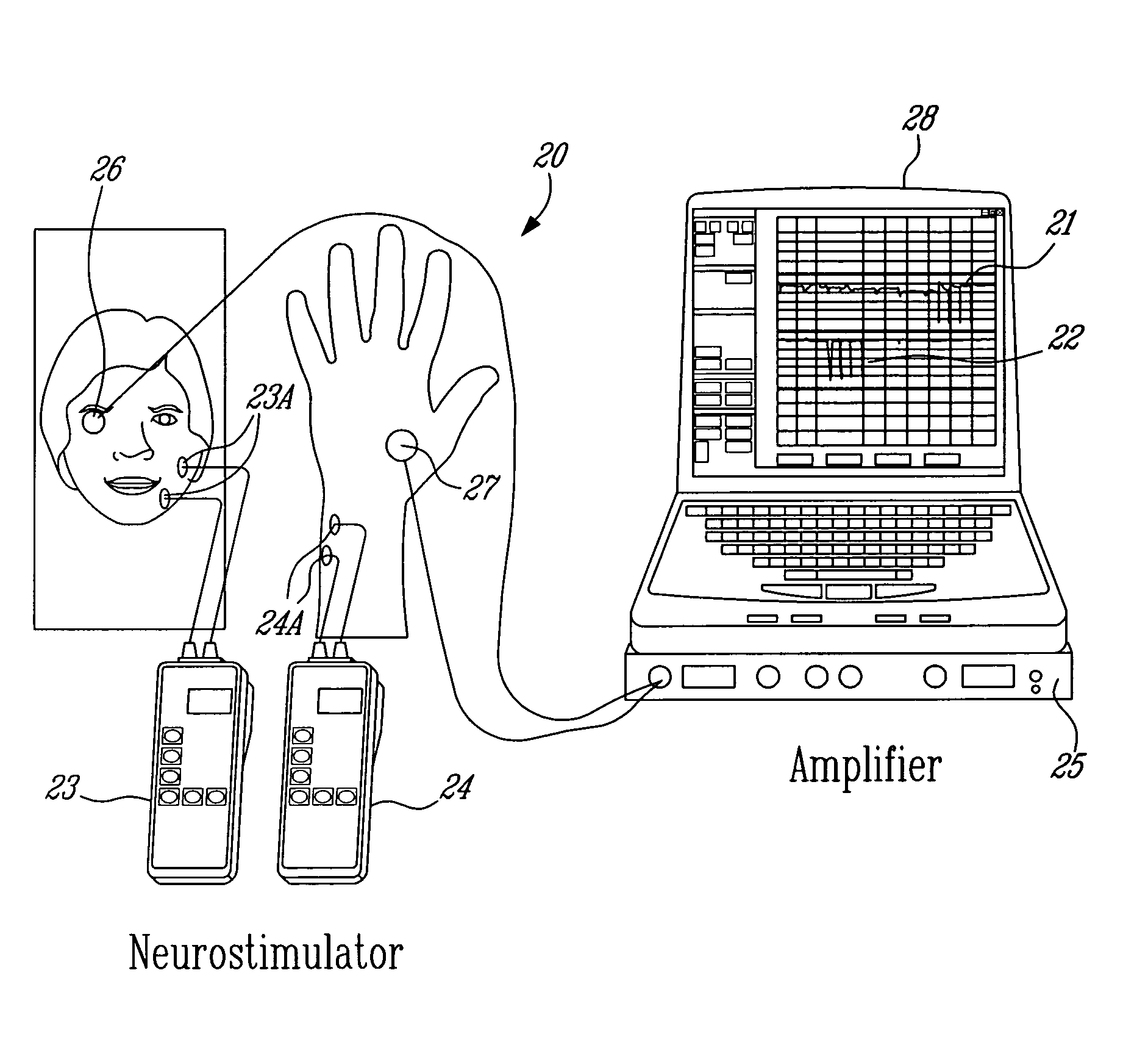
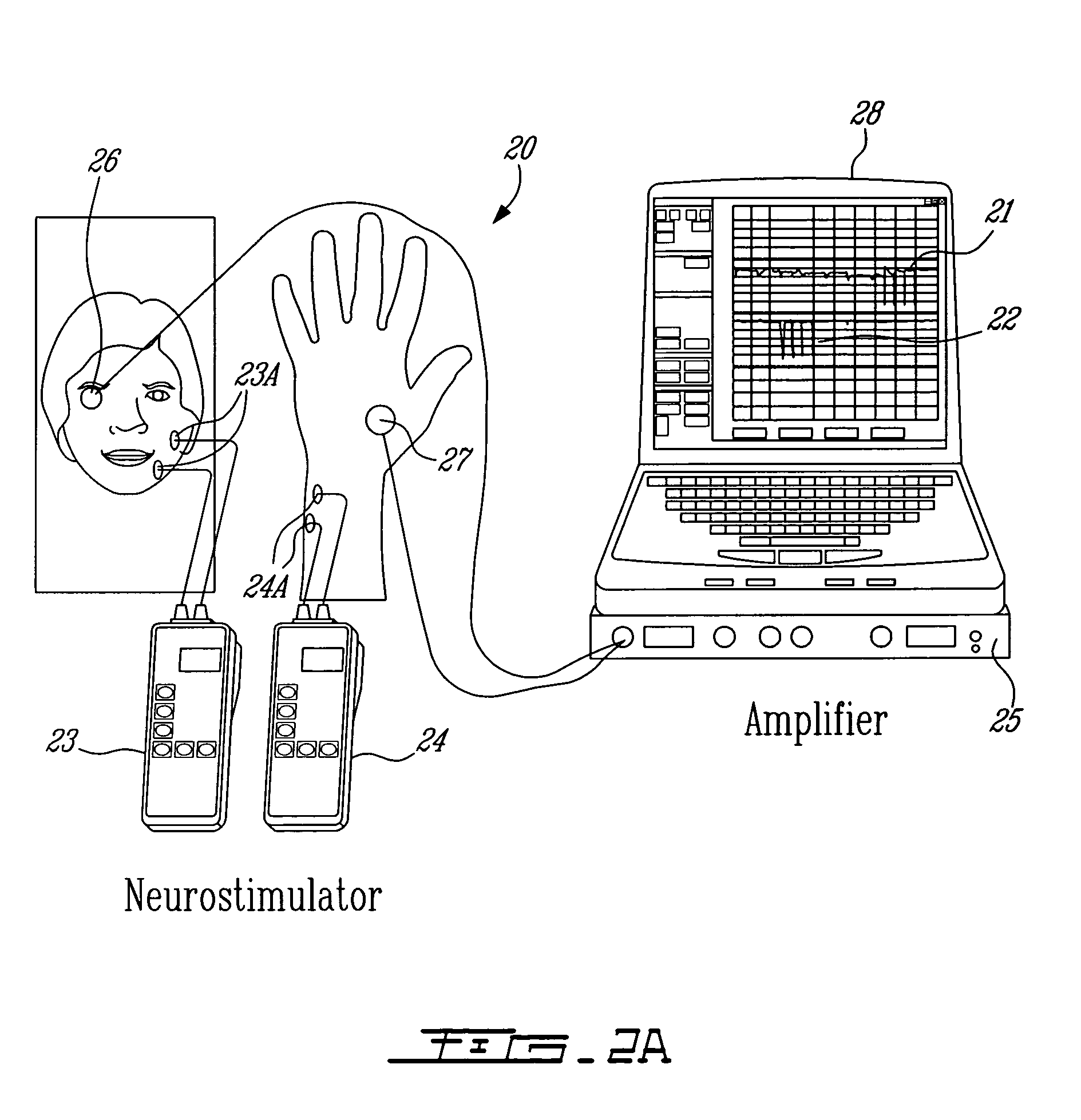
[0045]Muscle contraction creates pressure waveforms. Phonomyography is the detection of these pressure waveforms with a low frequency sensitive microphone acting as a pressure waveform sensor [9]. Detection of these pressure waveforms through phonomyography can be used to determine neuromuscular blockade [[4], [10] and [11]]. Since the amplitude of the sound waves detected at the microphone is a function not only of stiffness and tension of the muscle, but also of the distance and type of the tissue separating the muscle and the recording microphone, the position of the microphone in relation to the muscle and the monitored muscle affects the signal characteristics [9].

[0046]In the illustrative embodiments of the present invention, phonomyography is used as a method for monitoring neuromuscular blockade at all muscles of interest. It is believed that phonomyography could become a new standard of neuromuscular monitoring for research and clinical routine. Phonomyography has shown to ...


PUM


Abstract

A neuromuscular monitoring system comprises at least one neurostimulator to apply muscle-activating stimulation signals to a patient's body via at least one electrode, and at least one pressure waveform sensor to detect pressure waveform signals produced by a patient's muscle in response to the applied stimulation signals. The detected pressure waveform signals are processed and data related to these detected pressure waveform signals are displayed. A method for neuromuscular monitoring using pressure waveform sensors is also described.

Description

PRIORITY CLAIM[0001]The Application claims benefit of the U.S. Provisional Application Ser. No. 60 / 431,736 filed on Dec. 9, 2002 which is expressly incorporated herein, by reference.FIELD OF THE INVENTION[0002]The present invention relates to the use of phonomyography for performing neuromuscular monitoring.[0003]The present specification will refer to many references listed at the end of the disclosure, these references being herein incorporated by reference.BACKGROUND OF THE INVENTION[0004]Neuromuscular monitoring, the monitoring of muscle relaxation, is as essential as controlling blood pressure or heart rate during surgery. A patient who is extubated when still partially relaxed is at great risk of respiratory complications. Also, a patient incompletely relaxed during surgery can endanger the success of surgery.[0005]Since muscle relaxants are an important cost factor in anesthetic drug selection, neuromuscular monitoring also helps to titrate the exact dosing of muscle relaxant...

Claims


Application Information

Patent Timeline
Patent Type & Authority Patents(United States)
IPC IPC(8): A61N1/00A61B7/00A61B8/00A61N1/08
CPCA61B7/006A61N1/08A61B5/4821A61B2560/0443A61B5/7217
Inventor HEMMERLING, THOMASMATHIEU, PIERRE A.DONATI, FRANCOISTRAGER, GUILLAUMEGUARDO, ROBERTBABIN, DENISBOU-PHON, CHANG
Owner UNIV DE MONTREAL
Why Eureka
Try Eureka