Device for biopsy of tumors

a tumor and biopsy technology, applied in the field of breast cancer diagnosis and treatment, can solve the problems of difficult needle force into the tumor, tumors that are too tough to yield to suction and deformity, and devices that are not designed for resection, so as to prevent the destruction of tumor cells and reduce the dispersion of tumor cells

Inactive Publication Date: 2007-12-25
SCION MEDICAL
View PDF40 Cites 60 Cited by
  • Summary
  • Abstract
  • Description
  • Claims
  • Application Information

AI Technical Summary

Benefits of technology

[0008]The devices also include a coring apparatus with structures that permit the surgeon to core a sample of the tumor during the biopsy procedure. The coring apparatus is provided with a cannula that advances through a tumor to core a sample of the tumor. The coring apparatus is adapted for use with the probe. The probe is inserted into the cannula with the distal tip of the probe extending beyond the distal tip of the cannula. The surgeon can insert the devices into the body until the probe pierces the tumor. Coolant is directed to the distal tip of the probe to lightly cool the distal tip and the tumor. The lightly cooled distal tip adheres to the tumor cells. Once secured to the probe, the surgeon can core a sample of the tumor with the coring apparatus. After coring is complete, the surgeon can retract the device with the core sample. This method of biopsy prevents destruction of the tumor cells and reduces the dispersion of tumor cells to healthy cell areas.

Problems solved by technology

It is difficult to force a needle into these lesions because they are resistant to puncture and fairly mobile.
However, the tumor itself must be re-engaged after every coring operation, which entails substantial effort in relocation and confirmation that the target suspect mass has been engaged by the side aperture.
Tumors may be too tough to yield to the suction and deform as necessary to enter the side opening of the cannula.
Indeed, the device is not designed to perform resection with assurance that complete resection of a suspect mass has been accomplished.
Mechanical cutting and disruption of the tissue structure and cancer cell dispersion (that is, tearing of the tissue around the cancer and movement of the cancer cells amongst normal tissue) will result in unintentional delivery of cancer cells into healthy tissue adjacent the lesion.

Method used


Image

  • Device for biopsy of tumors
  • Device for biopsy of tumors
  • Device for biopsy of tumors

Examples


Embodiment Construction

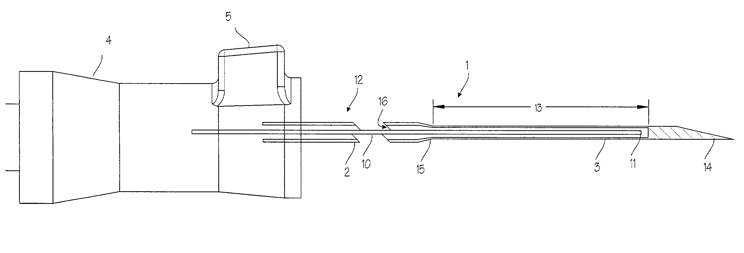
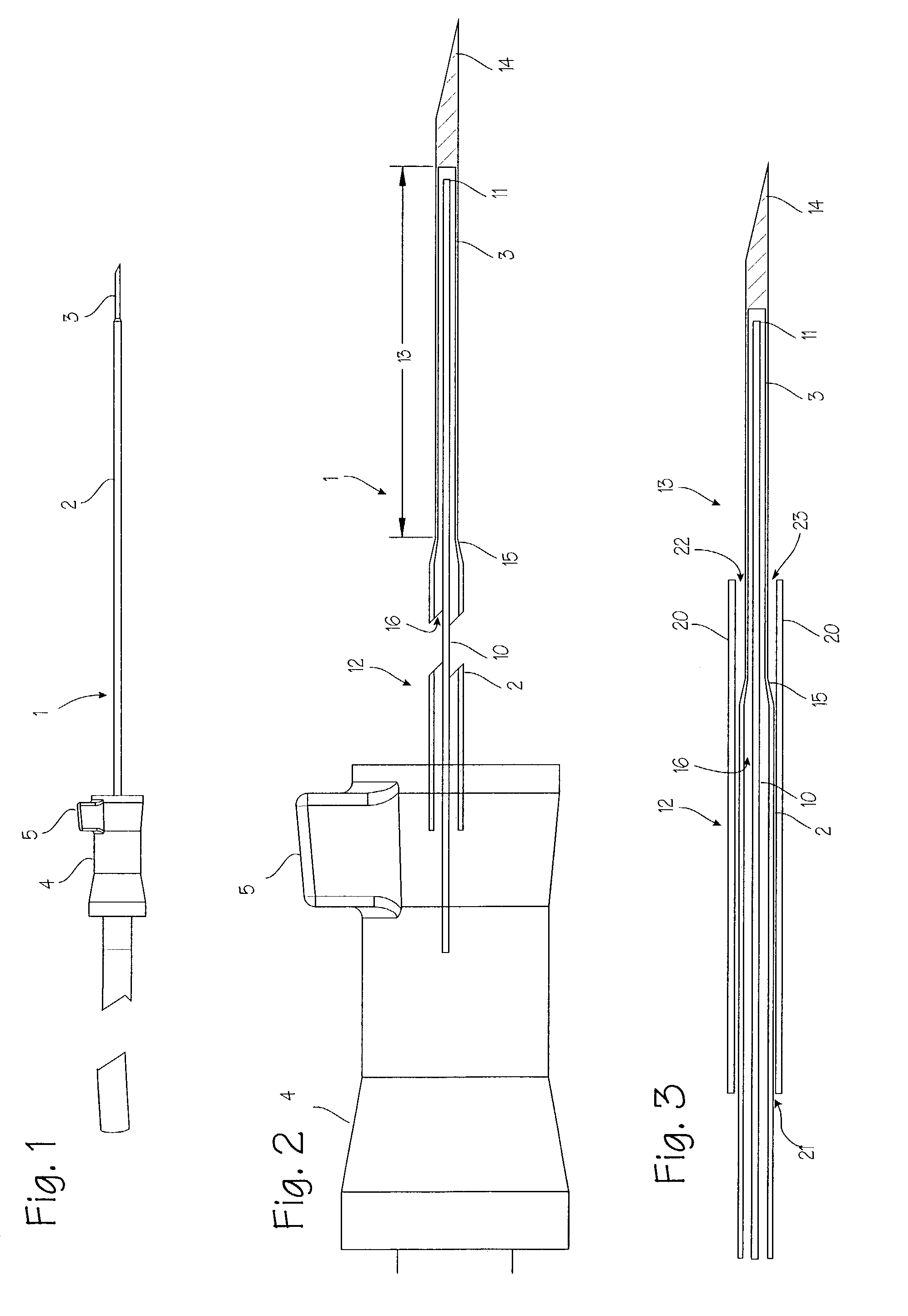
[0031]FIG. 1 illustrates an adhesion probe 1 for securing a breast tumor during a biopsy or resection procedure. This probe uses Joule-Thomson cooling or liquid nitrogen to create a lightly cooled region at the distal tip. This lightly cooled region adheres to a suspect lesion or tumor. The adhesion probe 1 comprises a long, slender yet rigid tube 2. A short rigid penetrating segment 3 extends distally from the distal end of the rigid tube, and a suitable handle 4 is mounted on the proximal end of the tube. The handle includes a quick release mechanism which is operable through quick release actuator 5.

[0032]FIG. 2 illustrates the adhesion probe 1 in cross section, showing the rigid tube 2, the distal penetrating segment 3 and the handle 4. A coolant inlet tube 10 passes through the handle and the rigid tube, extending to the distal end of the rigid tube, and terminating just proximal of the distal tip of the penetrating segment. The inlet tube has an orifice 11 at the distal end of...


PUM


Abstract

A device and method of use for securing and coring of tumors within the body during a biopsy of the tumor, specifically breast tumors. An adhesion probe for securing the tumor is described. The probe secures the tumor by piercing the tumor and providing a coolant to the distal tip to cool the tip. The cooled tip adheres to the tumor. A coring instrument adapted for cutting a core sample of the tumor is described. The instrument is provided with a cannula that can cut a core sample of the tumor. The instrument is adapted for use with the probe with the probe fitting within the cannula. The instrument can be used in conjunction with the probe to secure and core a sample of the tumor for biopsy.

Description

[0001]This application is a continuation application of U.S. application Ser. No. 09 / 847,931, filed May 3, 2001, now U.S. Pat. No. 6,551,255, which is a continuation-in-part application of U.S. application Ser. No. 09 / 690,321, filed Oct. 16, 2000, now U.S. Pat. No. 6,540,694.FIELD OF THE INVENTIONS[0002]The devices and method described below relate to the diagnosis and treatment of breast lesions, and more generally, to the diagnosis and treatment of tumors and lesions throughout the body.BACKGROUND OF THE INVENTIONS[0003]Biopsy is an important procedure used for the diagnosis of patients with cancerous tumors, pre-malignant conditions, and other diseases and disorders. Typically, in the case of cancer, when the physician establishes by means of procedures such as palpation, mammography or x-ray, or ultrasound imaging that suspicious circumstances exist, a biopsy is performed. The biopsy will help determine whether the cells are cancerous, the type of cancer, and what treatment shou...

Claims


Application Information

Patent Timeline
Patent Type & Authority Patents(United States)
IPC IPC(8): A61B10/00H04B7/00A61B10/02A61B18/02A61B19/00H04L12/24H04L12/28H04L12/56H04W36/16
CPCA61B10/0266H04L41/147H04L47/11H04L47/14A61B18/02A61B2010/0208A61B2018/0293A61B2018/0262H04W8/04
Inventor VAN BLADEL, KEVIN H.TANAKA, SHIGERUSTEWART, DAREN L.
Owner SCION MEDICAL
Why Eureka
Try Eureka