Tissue stabilizer having an articulating lift element

a tissue stabilizer and lift element technology, applied in the field of surgical instruments, can solve the problems of complicated completion of anastomosis, higher likelihood of surgical error in the formation of anastomosis, and forces alone can do little to optimize the visualization and presentation of target, and achieve the effect of facilitating the manipulation of the target coronary artery

Inactive Publication Date: 2008-02-05
MAQUET CARDIOVASCULAR LLC
View PDF273 Cites 78 Cited by
  • Summary
  • Abstract
  • Description
  • Claims
  • Application Information

AI Technical Summary

Benefits of technology

[0010]The present invention will be described for use during CABG surgery, but the invention is not limited thereto, and is contemplated to be useful for other surgical procedures as well. The present invention involves a tissue stabilizer having one or more stabilizer feet which are adapted to engage the heart tissue adjacent a target artery desired to be stabilized. In preferred embodiments of the present invention, the tissue stabilizer feet typically have a first foot portion which provides stabilization and a moveable portion which primarily facilitates the manipulation of the target coronary artery or local tissue surrounding the target coronary artery.
[0021]Another aspect of the present invention involves a device for stabilizing tissue within a patient's body for performing a surgical procedure on the tissue comprising a first stabilizer foot having a first tissue engaging surface, a second stabilizer foot having a second tissue engaging surface, and base member having a shaft mounted for rotation relative to the base member. The shaft preferably has at least one threaded portion. The first and second tissue engaging surfaces may extend generally perpendicular to the longitudinal axis of the shaft. At least one of the stabilizer feet may be operably associated with the threaded portion of the shaft such that rotation of the shaft causes one of the stabilizer feet to move relative to the other.
[0026]In a preferred embodiment, the tissue stabilizer has first and second stabilizer feet, each having a first portion and a second portion coupled to said first portion and moveable relative to said first portion. The method preferably includes the step of adjusting one or both of the stabilizer feet relative to each other to ensure a good fit against the surface of the heart. The first portion of each stabilizer foot may then be engaged with the surface of the heart on opposite sides of the coronary artery. An arteriotomy having a first side and a second side may be created in the coronary artery. In the preferred method, the first side is positioned relative to the second side by engaging one or both of the second portions of the stabilizer feet with the heart and moving one or both relative to the first portions. The method may also include the step of manipulating the first and second stabilizer feet relative to one another to improve presentation of the arteriotomy.

Problems solved by technology

Forming an anastomosis between two vessels in this manner is a particularly delicate procedure requiring the precise placement of tiny sutures in the tissue surrounding the arteriotomy in the coronary artery and in the source or graft vessel so that the two may be sutured together.
While a properly stabilized vessel will usually exhibit acceptable visualization, some operations and tissue geometries can distort the tissue surrounding the coronary artery or the coronary artery itself in a manner which complicates the completion of the anastomosis.
In such cases, the stabilization forces alone can do little to optimize the visualization and presentation of the target artery.
When the arteriotomy is not optimally presented, there is a higher likelihood of incurring a surgical error in the formation of the anastomosis.
If the vessel is flattened, for example, it becomes more likely for the surgeon to accidentally catch the back wall of the vessel with the curved suture needle as the suture is placed in the tissue surrounding the arteriotomy.
When the vessel is compressed side to side, it becomes more difficult to catch only the desired side of the arteriotomy without also catching the other side.
The surgeon must be careful to only manipulate the vessel at the outside edges of the incision as manipulation to the interior of the vessel wall may cause damage to the soft intimal layer of the vessel leading to scarring and often late restenosis.
Even in the best of circumstances this manual manipulation of the vessel to facilitate each suture placement is tedious, time consuming, and increases the likelihood of vessel damage.
This problem will become even more magnified as surgeons move to manual, computer-assisted, and robotic endoscopic procedures in which the surgeons will be attempting to complete anastomotic procedures in remote and difficult places.
Delicate manipulation of the vessel walls while suturing becomes increasingly difficult as the surgeon becomes further removed from the surgical site by longer instruments, the size of the surgical site decreases leaving inadequate space to accommodate multiple instruments and the access incisions become smaller thus limiting instrument maneuverability.

Method used


Image

  • Tissue stabilizer having an articulating lift element
  • Tissue stabilizer having an articulating lift element
  • Tissue stabilizer having an articulating lift element

Examples


Embodiment Construction

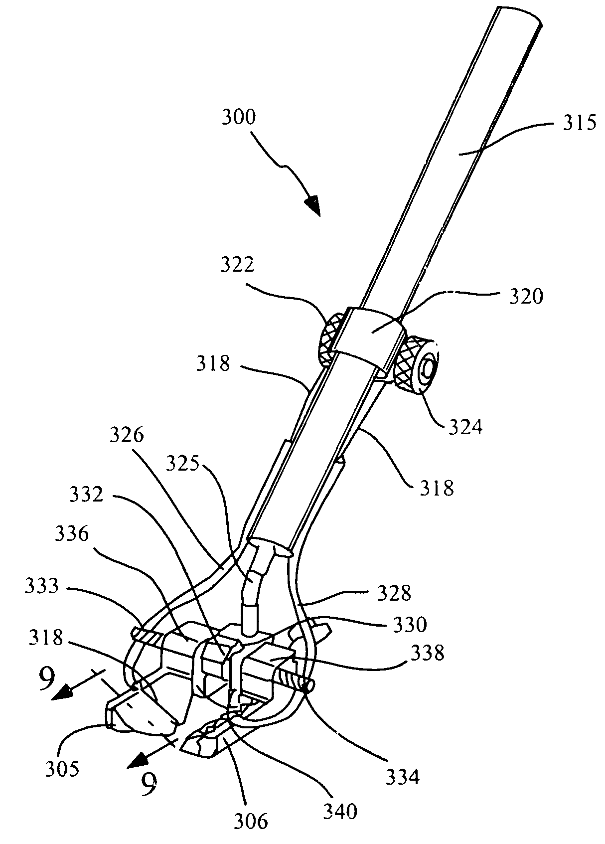
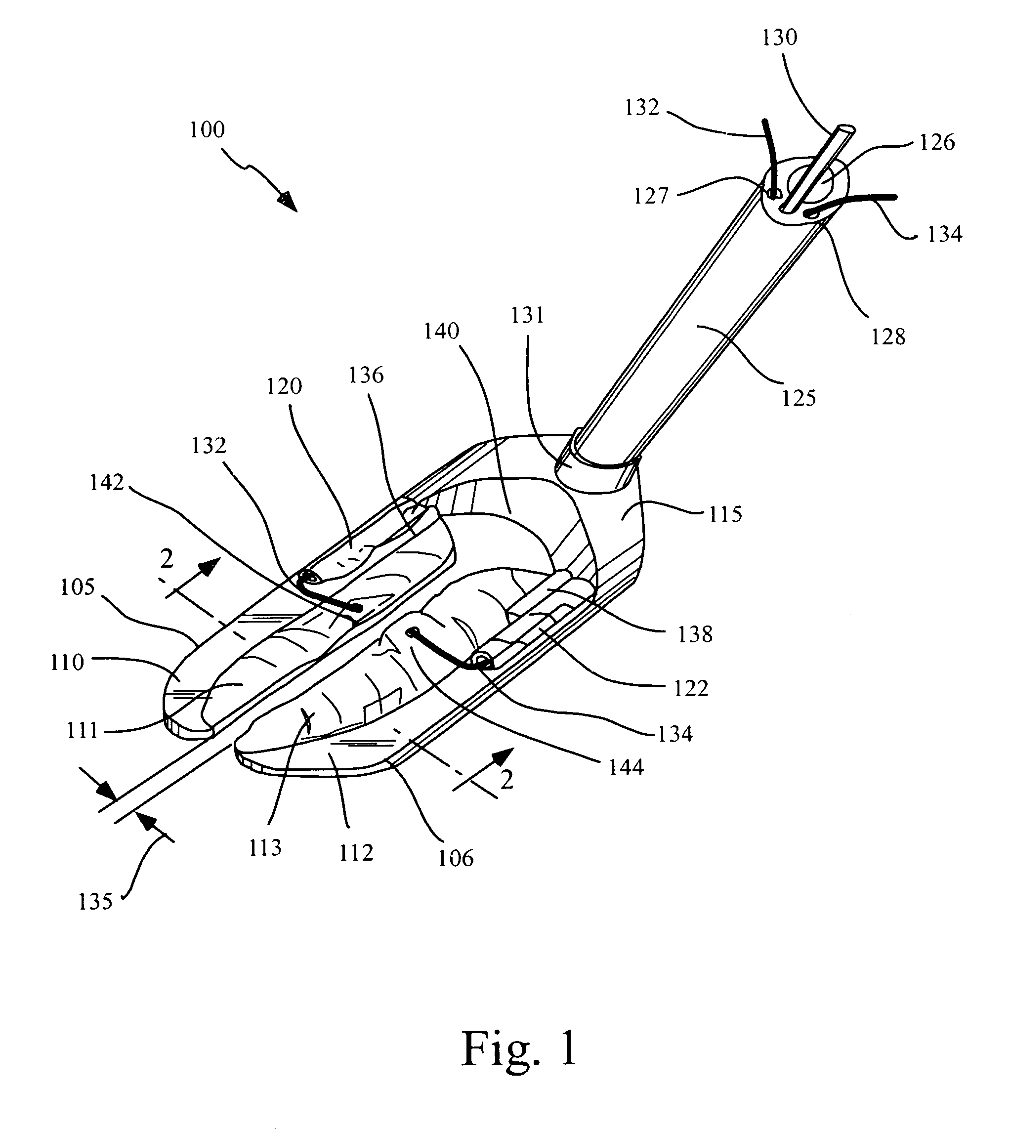
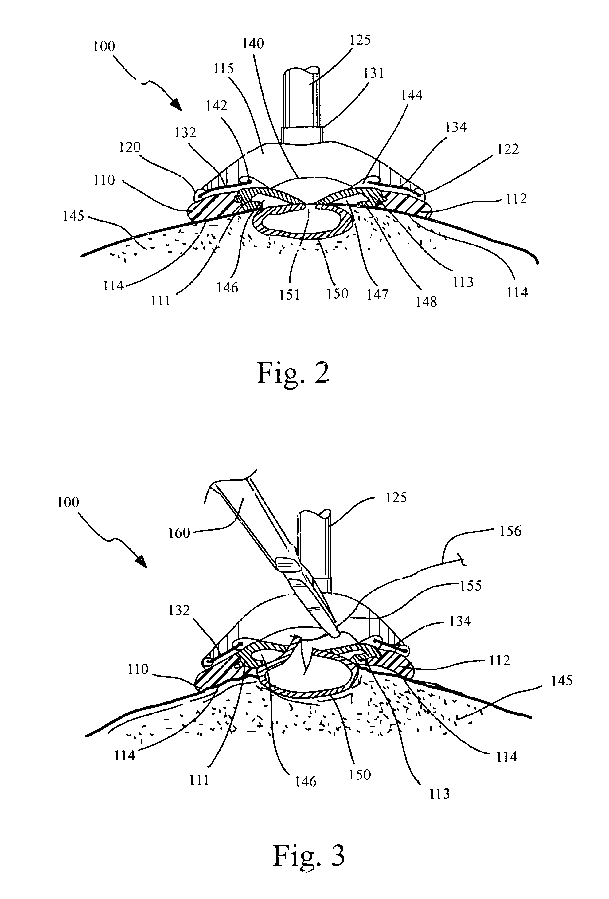
[0043]The present invention involves devices and methods for stabilizing tissue during a surgical operation. The devices described herein may be used in a wide variety of surgical applications that require a tissue structure to be stabilized or immobilized to provide a substantially stable and motionless surgical field on which a surgical procedure can be performed. By way of example only, the preferred embodiments described in detail below are directed to the stabilization of a portion of the heart to facilitate a surgical procedure on or within the heart, such as a coronary artery bypass graft (CABG) procedure.

[0044]Although the devices and methods of the present invention may be applied to conventional stopped-heart and beating heart procedures, they are preferably used to stabilize the beating heart during a CABG operation which has been specially developed to facilitate completion of an anastomosis, typically between a target coronary artery and a bypass graft or source artery,...


PUM


Abstract

Devices and methods are disclosed for stabilizing tissue within a patient's body during a surgical operation to provide a relatively motionless surgical field, such as during a coronary artery bypass graft procedure. The devices include tissue stabilizers which engage and provide stabilization to a targeted area of tissue and further have the ability to engage and manipulate some portion of tissue within or adjacent the targeted area to improve the surgical presentation of that portion of tissue. The tissue stabilizer typically has one or more stabilizer feet which have a first foot portion configured to provide stabilization to the targeted tissue and a second foot portion moveable relative to the first foot portion for manipulating a portion of tissue to improve the surgical presentation.

Description

[0001]This application is a continuation of application Ser. No. 09 / 398,535, filed Sep. 16, 1999, now U.S. Pat. No. 6,406,424, which is incorporated herein by reference in its entirety and to which application we claim priority under 35 USC § 120.FIELD OF THE INVENTION[0002]The present invention relates generally to surgical instruments, and more particularly to devices and methods for stabilizing and manipulating tissue during surgery. The tissue stabilizers described herein are particularly useful for stabilizing the beating heart during coronary artery bypass graft surgery. The field of the invention is cardiac tissue stabilizers having specially designed moveable portions.BACKGROUND OF THE INVENTION[0003]Certain surgical procedures require the surgeon to perform delicate operations on tissues within the body that are moving or otherwise unstable. For example, a large and growing number of surgeons are routinely performing successful coronary artery bypass graft surgery on the be...

Claims


Application Information

Patent Timeline
Patent Type & Authority Patents(United States)
IPC IPC(8): A61B1/32A61F2/00A61B17/02
CPCA61B1/32A61B17/02A61B2017/0243A61B2017/306
Inventor WILLIAMSON, IV, WILLIAM P.SPENCE, PAUL A.ORTIZ, MARKKELLER, GEORGE A.GREEN, II, HARRY LEONARD
Owner MAQUET CARDIOVASCULAR LLC
Why Eureka
Try Eureka