Methods and devices for draining fluids and lowering intraocular pressure

a technology of intraocular pressure and draining fluid, applied in the field of medicine and surgery, can solve the problems of abnormally high intraocular pressure, inferior and superior damage to produce characteristic arcuate defects, and the lamina cribosa is not well supported, so as to prevent unwanted backflow of fluid and prevent or deter clogging of the shunt devi

Active Publication Date: 2008-04-08
QUIROZ MERCADO HUGO +1
View PDF11 Cites 58 Cited by
  • Summary
  • Abstract
  • Description
  • Claims
  • Application Information

AI Technical Summary

Benefits of technology

[0014]Further in accordance with the invention, there is provided a shunt device for draining fluid from the posterior chamber of the eye into the optic nerve or subarachnoid space. Such shunt device comprises a tube having a proximal end, a distal end and a lumen extending longitudinally therethrough, a substantially tissue penetrating tip on the distal end of the tube, a plurality of openings formed at or near the distal end of the tube to allow fluid to drain out of the lumen of the tube and at least one tissue engaging member configured to allow the shunt device to be advanced, tip member first, into tissue but to engage said tissue in such a manner as to subsequently deter retraction of the shunt out of the tissue. Optionally, the shunt device may additionally include a pressure control and / or one-way valve to control the magnitude of the pressure head required to cause fluid to drain from the eye through the shunt device and / or to prevent unwanted backflow of fluid into the eye through the shunt device. Also, optionally, the shunt device may comprise a shielding member, such as a semi-permeable membrane, to prevent or deter clogging of the shunt device by foreign matter and or tissue in-growth.

Problems solved by technology

Glaucoma is typically, but not always, accompanied by abnormally high intraocular pressure.
The lamina cribosa is not well supported superiorly and inferiorly at the disk and, as a result, initial damage occurs superiorly and inferiorly to produce the characteristic arcuate defects.
Presently the use of topically applied glaucoma medications consisting of mainly beta blockers, prostaglandin analogues, alpha-2 agonists, and carbonic anhydrase inhibitors are short acting, prone to deleterious side effects, prone to compliance issues, and must be used for life.
Also, at present, the use of argon laser trabeculoplasty as a means for treating glaucoma is limited in clinical response, lasts only approximately 1-2 years, and is limited by the number of applications per eye.
However, trabeculectomy procedures can be technically difficult, frought with early hypotony, late failure, and high rate of endophthalmitis leading to permanent loss of the eye.

Method used


Image

  • Methods and devices for draining fluids and lowering intraocular pressure
  • Methods and devices for draining fluids and lowering intraocular pressure
  • Methods and devices for draining fluids and lowering intraocular pressure

Examples


Embodiment Construction

[0025]The following detailed description and the accompanying drawings are provided for the purpose of describing certain non-limiting examples or embodiments of the invention only. This detailed description is not intended to describe all possible examples and embodiments of the invention and, thus, shall not limit the scope of the claimed invention in any way.

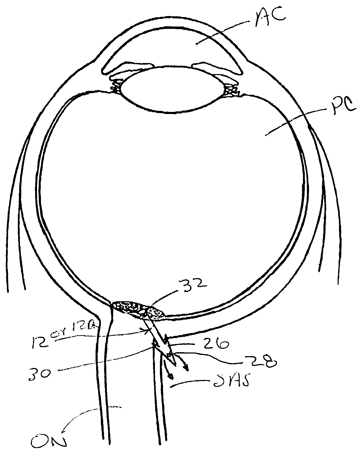
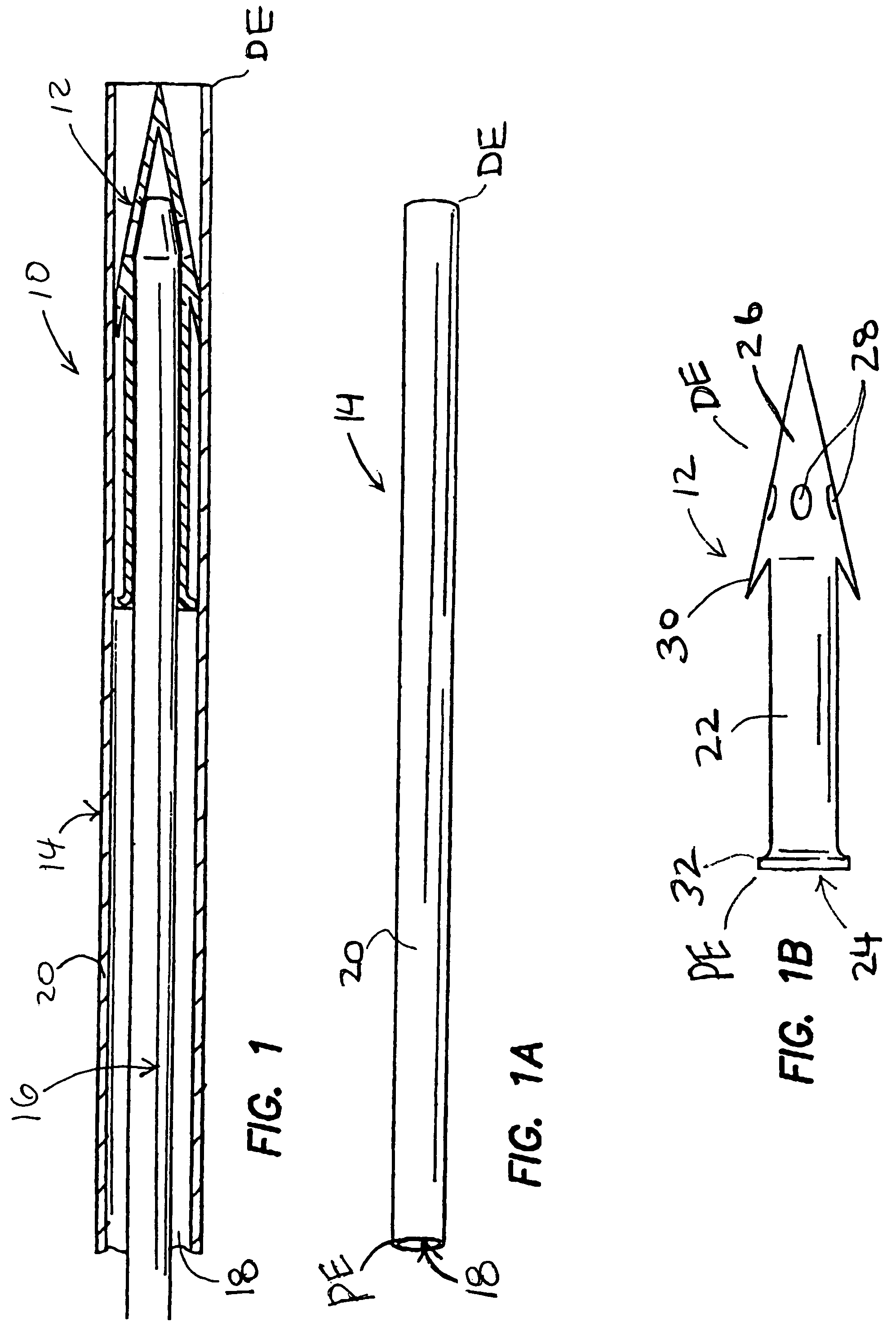
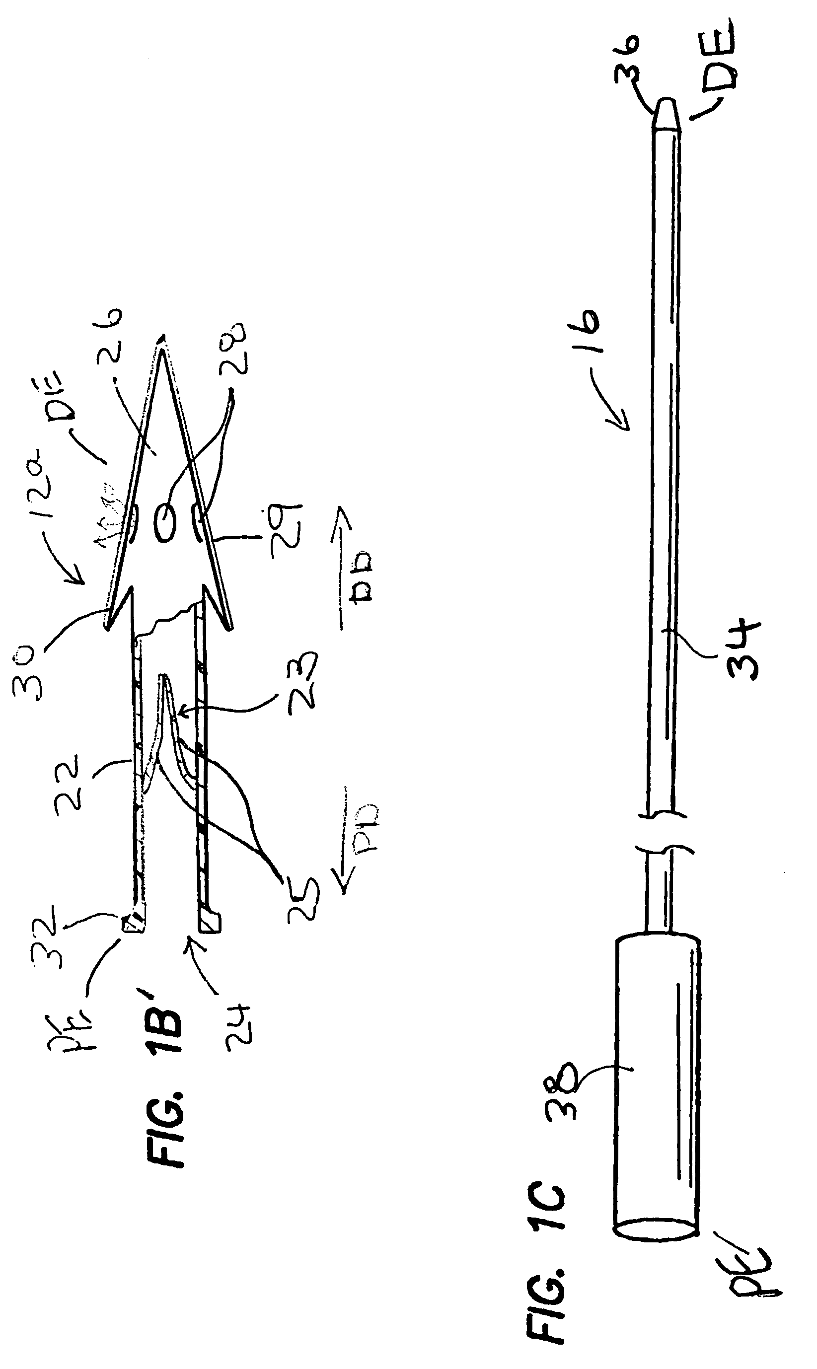
[0026]FIGS. 1-1C show one embodiment of a system 10 for implantation of a shunt device 12 in accordance with the present invention. This system 10 generally comprises the shunt device 12, a cannula 14, and a pusher 16. As shown in FIG. 1, the shunt device 12 is initially positioned within the lumen 18 of the cannula 14 and the pusher 16 is positioned in the lumen 18 of the cannula 14 behind the shunt device 12 such that the pusher 16 may be used to push the shunt device 12 out of the distal end DE of the of the cannula 14. One way of performing this shunt-expulsion procedure is shown in FIG. 2A and is explained in detail here...


PUM


Abstract

Methods, devices and systems for draining fluid from the eye and / or for reducing intraocular pressure. A passageway (e.g., an opening, puncture or incision) is formed in the lamina cribosa or elsewhere to facilitate flow of fluid from the posterior chamber of the eye to either a) a subdural location within the optic nerve or b) a location within the subarachnoid space adjacent to the optic nerve. Fluid from the posterior chamber then drains into the optic nerve or directly into the subarachnoid space, where it becomes mixed with cerebrospinal fluid. In some cases, a tubular member (e.g., a shunt or stent) may be implanted in the passageway. A particular shunt device and shunt-introducer system is provided for such purpose. A vitrectomy or vitreous liquefaction procedure may be performed to remove some or all of the vitreous body, thereby facilitating creation of the passageway and / or placement of the tubular member as well as establishing a route for subsequent drainage of aqueous humor from the anterior chamber, though the posterior chamber and outwardly though the passageway where it becomes mixed with cerebrospinal fluid.

Description

RELATED APPLICATION[0001]This application claims priority to U.S. Provisional Patent Application No. 60 / 447,999 filed on Feb. 18, 2003, the entirety of which is expressly incorporated herein by reference.FIELD OF THE INVENTION[0002]This invention relates generally to medicine and surgery, and more particularly to methods and devices for lowering intraocular pressure in human or veterinary patients.BACKGROUND OF THE INVENTION[0003]In normal adults the ocular globe is approximately spherical, with a diameter averaging 24.5 mm. The cornea is a transparent tissue inserted into the sclera at the limbus, the anterior chamber is behind the cornea. The iris is the anterior extension of the ciliary body, it presents as a flat surface with a centrally situated round aperture, the pupil. The iris lies in contiguity with the anterior surface of the lens, dividing the anterior chamber from the posterior chamber, each of which contains aqueous humor. The lens is a biconvex, avascular, colorless a...

Claims


Application Information

Patent Timeline
Patent Type & Authority Patents(United States)
IPC IPC(8): A61M5/00A61B17/08A61B19/00A61FA61F2/14A61F9/007
CPCA61F9/00781
Inventor QUIROZ-MERCADO, HUGOKARAGEOZIAN, HAMPAR L.
Owner QUIROZ MERCADO HUGO
Why Eureka
Try Eureka