

Serum macrophage migration inhibitory factor (MIF) as marker for prostate cancer
a prostate cancer and migration inhibitor technology, applied in the field of prostate cancer diagnosis and prognosis, can solve the problem that the biopsy process often does not recover useable tissue, and achieve the effect of aggressive treatment and aggressive diseas
- Summary
- Abstract
- Description
- Claims
- Application Information
AI Technical Summary
Benefits of technology
Problems solved by technology
Method used
Image

Examples
example 1
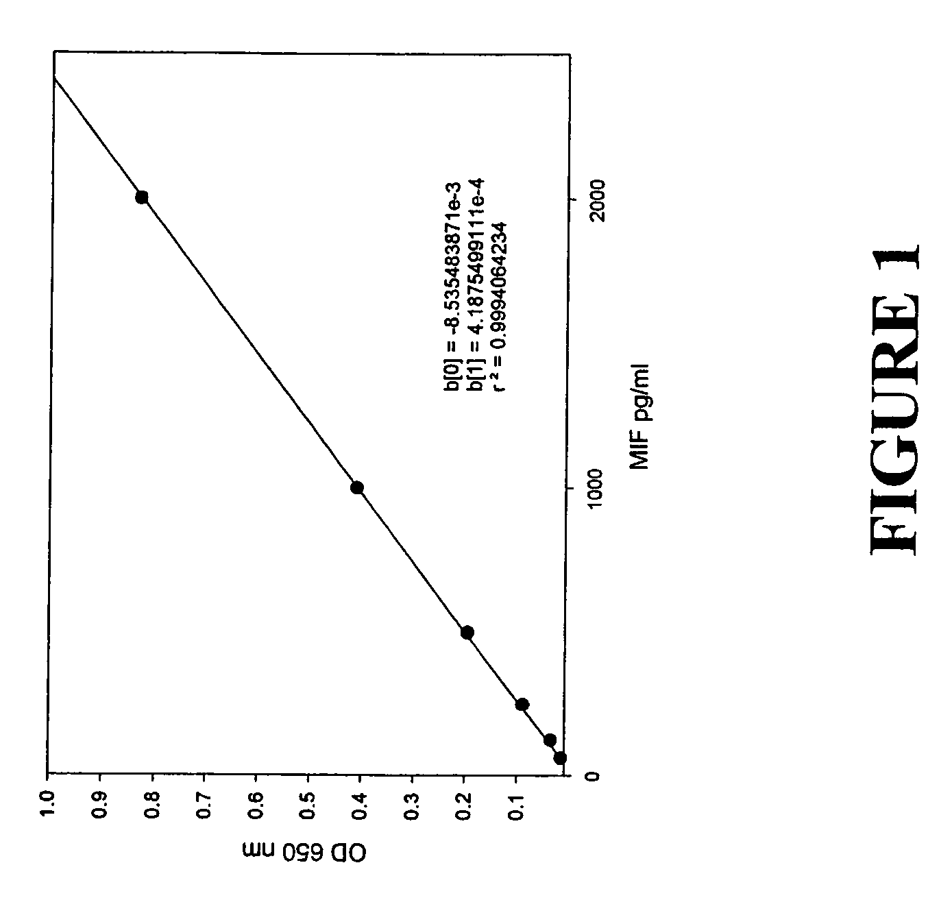
ELISA
[0037]A MIF-specific ELISA assay was developed, based on the capture of MIF by immobilized monoclonal anti-MIF antibody followed by detection with goat polyclonal anti-MIF affinity purified IgG. This assay was performed as follows:[0038]1. ELISA plate wells were coated with 2 μg / ml monoclonal antibody in PBS (100 μl / well); immediately following preparation of working monoclonal antibody (MAb) solution 100 μl aliquots of working antibody solution are transferred to each well of an ELISA plate. Plates are sealed and incubated overnight at 4° C.[0039]2. ELISA plate wells are washed. This process involves “flipping” the contents of the plate into a sink and rapping the plate three times on a wad of paper towels placed on top of a folded towel. Wells were the washed by filling the wells individually with wash buffer using a squeeze bottle, followed by removal of contents by “flipping” and drying on paper towels. The wash process is repeated for a total of four washes. At the end of ...
example 2
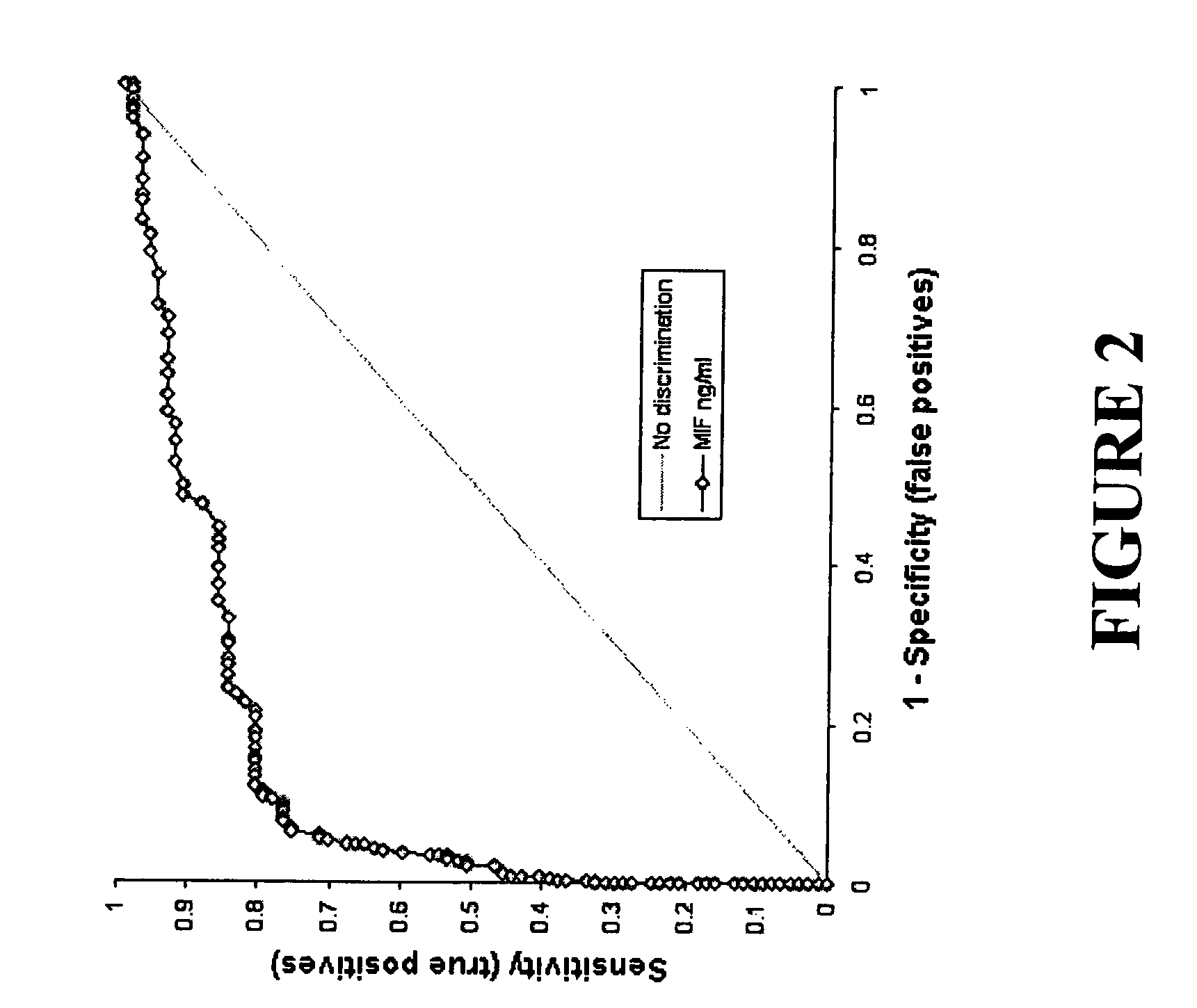
Serum MIF and Prostate Pathology
[0050]Four pathological groups were defined: benign prostatic hyperplasia (BPH), CaP, high-grade prostate intraepithelial neoplasia (HGPIN), and none available (patients without listed diagnosis or prostate pathology report). The mean serum MIF was determined for each group and is summarized in the following table:
[0051]
TABLE 1Serum MIF and Prostate PathologyProstate pathologySample size (n)Serum MIF (mean ± SE)None available3571.9 ± 0.28Benign prostatic hyperplasia842.7 ± 0.58CaP616.8 ± 0.87High grade PIN710.1 ± 3.35
[0052]Using serum from patients tested for thyroid stimulating hormone concentrations (n=14) as a control a pair wise Dunn's multiple comparison test of Krudkal-Wallis ANOVA on ranks identified significant difference in serum MIF levels between the control and high grade PIN, as well as control and CaP (P<0.001). These data suggest a correlation between CaP and elevated serum MIF.
[0053]Serum MIF concentrations were found not to correlate...
example 3
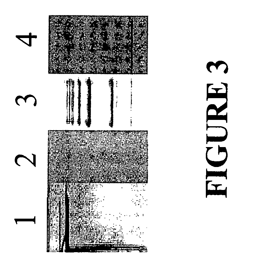
Detection of MIF Protein in Prostate Tissue
[0057]Previous experiments identified MIF as being localized within prostate epithelial cells (Meyer-Siegler (1998), Diagnostic and Molecular Path. 7: 44-50). Analysis of MIF staining within prostate tissue using the biopsy samples (same as used for serum analysis, Table 2) demonstrated an association between MIF immunoperoxidase staining and prostate tumor Gleason score (n=61, PProstate 39: 159-165). These authors suggest that this finding may be the consequence of changes in MIF synthesis or the result of an enhanced and / or altered secretion by tumor cells into the surrounding stroma (Arcuri et al.). Interestingly, the aforementioned study did not find significantly reduced intracellular MIF concentration in prostate tumor cells after patients underwent combined endocrine treatment (Arcuri et al.). These results and those showing elevated serum MIF in some CaP patients who have undergone endocrine treatment, which has reduced serum PSA to...
PUM
| Property | Measurement | Unit |
|---|---|---|
| Density | aaaaa | aaaaa |
| Density | aaaaa | aaaaa |
| Density | aaaaa | aaaaa |
Abstract
Description
Claims
Application Information