Methods and compositions in breast cancer diagnosis and therapeutics
- Summary
- Abstract
- Description
- Claims
- Application Information
AI Technical Summary
Benefits of technology
Problems solved by technology
Method used
Image
Examples
example 1
Materials and Methods—Sample Preparation and Nucleotide Sequence Analysis
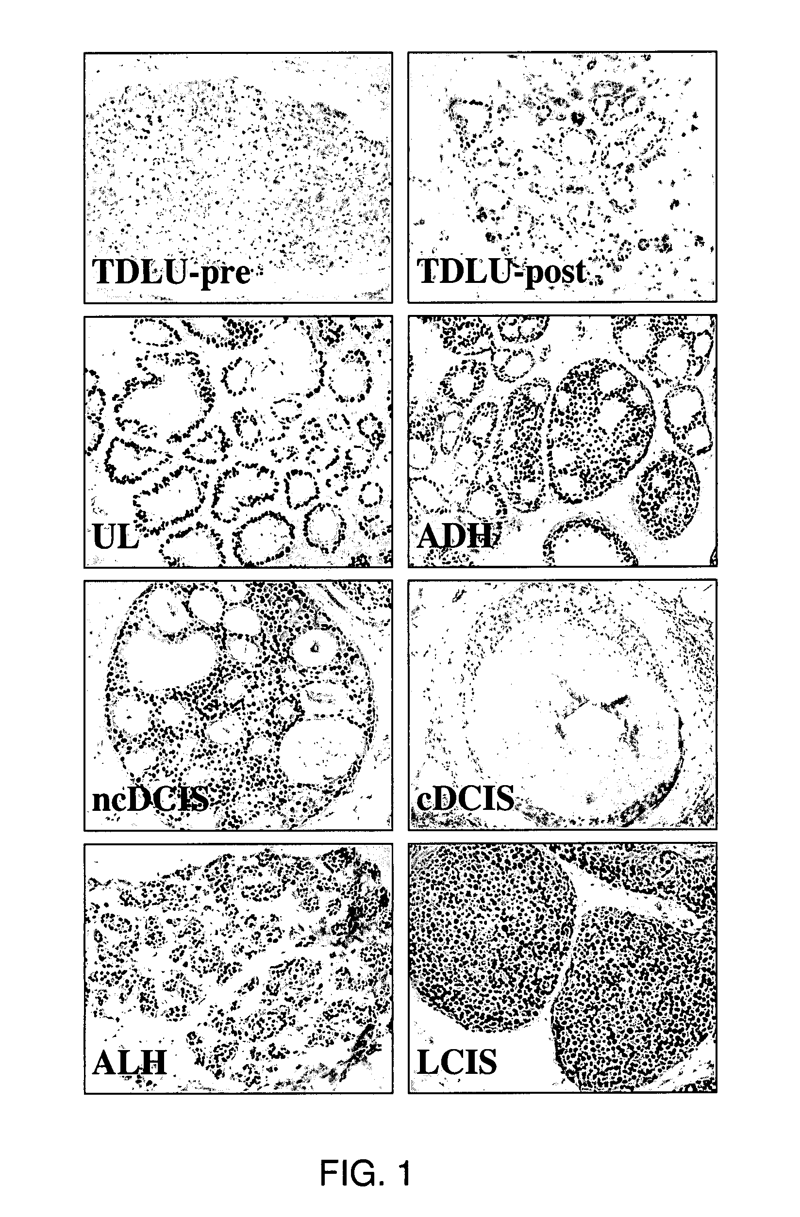
[0330]Histologic slides from archival, clinical specimens were screened microscopically for evidence of hyperplasia. Microdissection of specimens was performed on 55 samples using serial sections from formalin-fixed, paraffin-embedded tissue blocks as described (O'Connell et al., 1999). Briefly, alternative 3- and 10 um-thick sections were cut from the blocks and float mounted on glass slides. The 3-um-thick slides were stained with hematoxylin-eosin and examined under the light microscope to locate regions of normal and hyperplastic tissues; and these areas outlined with a felt-tipped pen. The marked slide was then used as a template to guide manual microdissection from the corresponding regions of the unstained 10-um-thick sections. It was possible to obtain distant normal tissue from 4 of the patients with hyperplasia. A skilled artisan recognizes that there are a variety of methods to isolate desired cells ...
example 2
Materials and Methods-Stable Transfection and Cell Growth Analyses
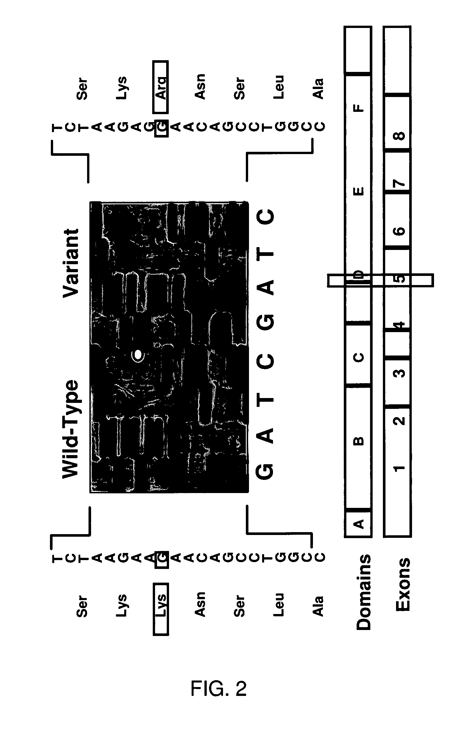
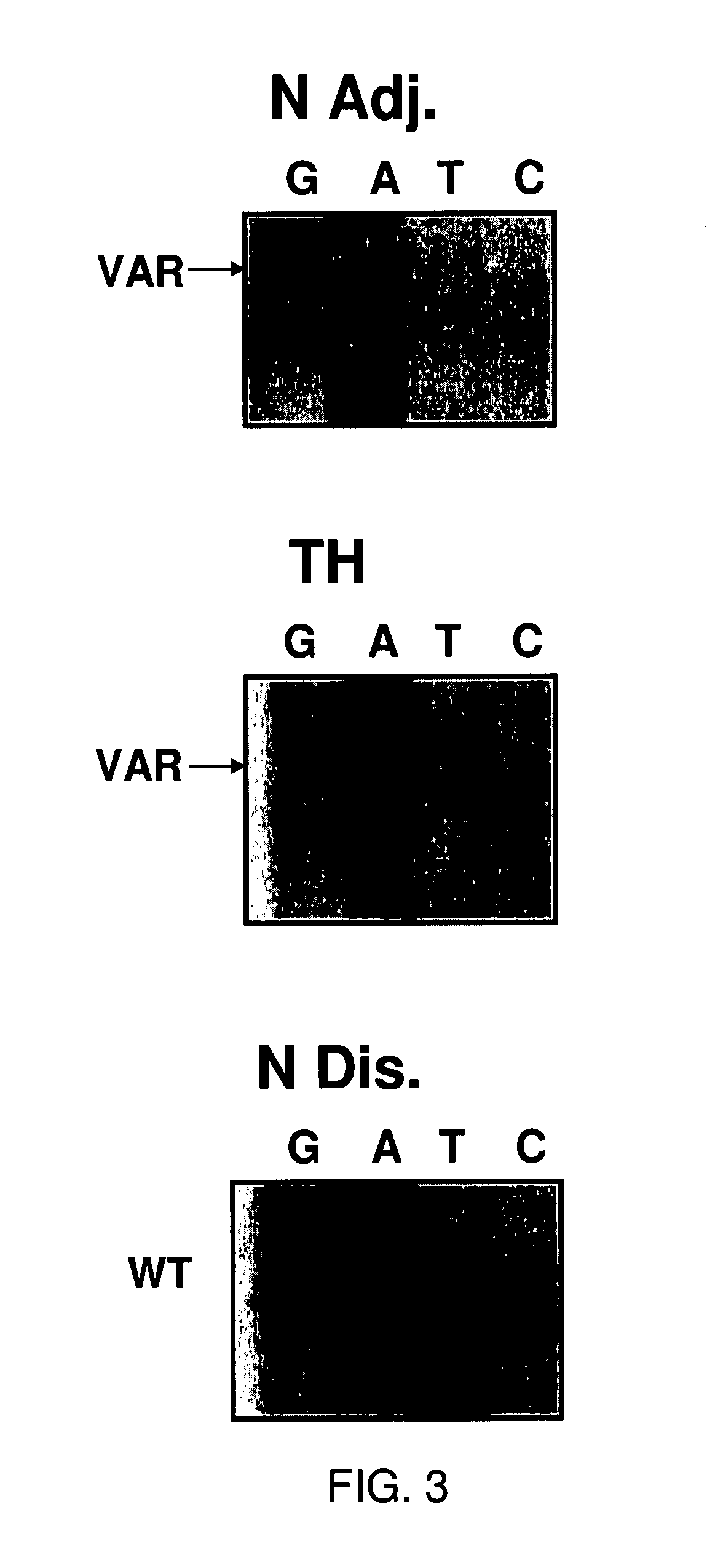
[0332]The WT ER expression construct was prepared in the pcDNAI vector as described previously (Fuqua et al., 1995). Site directed mutagenesis of this construct was then utilized to generate the A908G transition and the entire coding sequence of ER was verified by dideoxysequence analysis in this clone. The generation of stable transfectants was performed as described by Oesterreich et al. (1993) using cotransfection with the G418-selectable expression vector pSVneo at a ration of 25:1 with the ER plasmids into MCF-7 breast cancer cells. To analyze for expression of both WT or Var sequences, Western blot analyses were performed using the 6F11 antibody (DaKO). Two to three-fold elevated levels of total ER protein were detected in the two WT ER and the three Var clones. In addition, RT / PCR amplification of cDNA from the transfectants (Fuqua et al., 1991) followed by dideoxysequence analysis confirmed that exogenous WT a...
example 3
Statistical Methods
[0333]After taking logarithms to stabilize within group variances, as determined to be appropriate by Box-Cox analysis (Box and Cox, 1996), one-way analysis of variance was used to detect estrogen dose-related differences in growth on Day 8 (i.e. 0 versus 10−12 M versus 10−11 M versus 10−9 M), and to detect differences among estrogen doses (10−12 M versus 1011 M versus 10−9 M). The Student-Newman-Keuls multiple range test (a=0.02) was used to determine which doses were different from each other. Analyses were preformed using SAS (V6.12, SAS Institute, Cary, N.C.).
PUM
| Property | Measurement | Unit |
|---|---|---|
| Fraction | aaaaa | aaaaa |
| Fraction | aaaaa | aaaaa |
| Fraction | aaaaa | aaaaa |
Abstract
Description
Claims
Application Information
 - R&D
- Intellectual Property
- Life Sciences
- Materials
- Tech Scout
- Unparalleled Data Quality
- Higher Quality Content
- 60% Fewer Hallucinations
Browse by: Latest US Patents, China's latest patents, Technical Efficacy Thesaurus, Application Domain, Technology Topic, Popular Technical Reports.
© 2025 PatSnap. All rights reserved.Legal|Privacy policy|Modern Slavery Act Transparency Statement|Sitemap|About US| Contact US: help@patsnap.com


