Imaging elastic scattering spectroscopy

Active Publication Date: 2008-09-23
SPECTRAL MOLECULAR IMAGING
View PDF41 Cites 33 Cited by
  • Summary
  • Abstract
  • Description
  • Claims
  • Application Information

AI Technical Summary

Benefits of technology

[0016]The present invention provides a non-invasive technique for the early detection of cancer and other abnormal tissue. The present invention allows f

Problems solved by technology

It is therefore quite likely that a problem area, particularly one in an early stage of abnormality, may be missed completely.
Another serious drawback of this method of treatment is that since the identification of abnormality must await the pathology report, surgical removal of the abnormal tissue often must be performed during a separate procedure (and sometimes even by successive iterations), thereby increasing risk to the patient, and inconvenience and cost for both the patient and the physician.
Although numerous groups are working on the development of fluorescence-based systems for cancer diagnosis, to date only one device has reached the commercial

Method used


Image

  • Imaging elastic scattering spectroscopy
  • Imaging elastic scattering spectroscopy
  • Imaging elastic scattering spectroscopy

Examples


Embodiment Construction

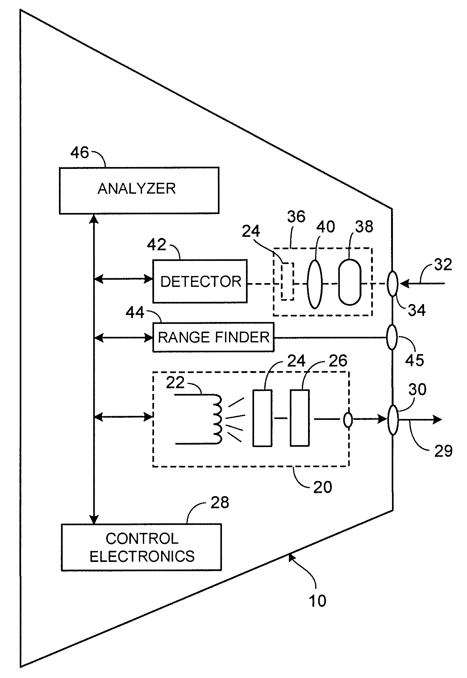
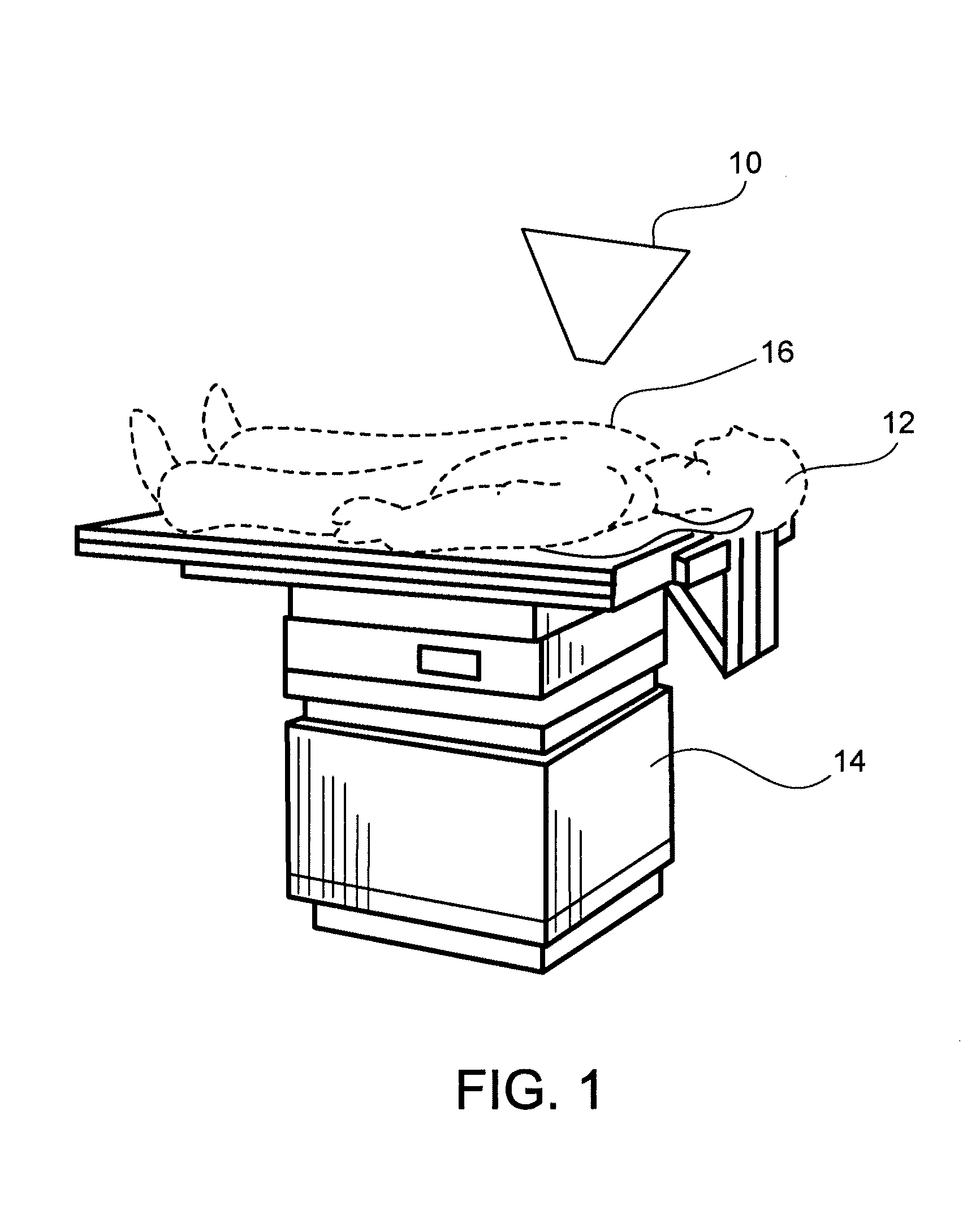
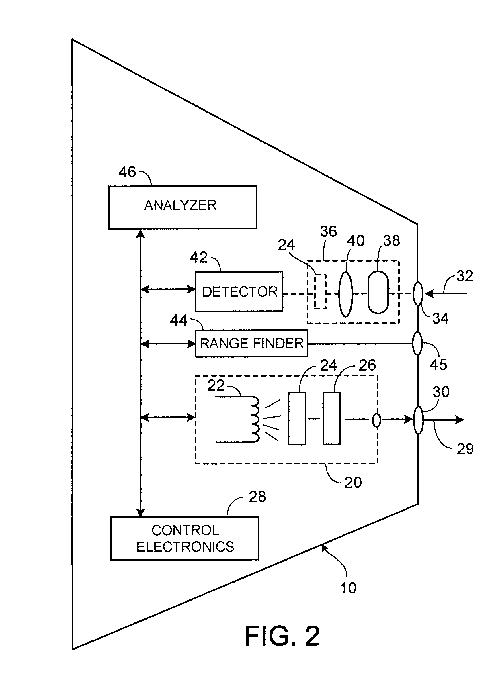
[0025]One embodiment of an apparatus 10 constructed according to the teachings of the present invention and useful for screening for abnormal cells is illustrated in FIG. 1. In FIG. 1, a patient 12 is positioned on an examining table 14. A target 16 is examined by apparatus 10 as will be described in detail below. Those of ordinary skill in the art will recognize that target 16 is meant to be exemplary and not limiting.

[0026]Most internal and external surfaces of the body are covered with a layer of cells known as the epithelium. One of the more common types of epithelial tissue is known as the “columnar epithelium”, in which a single layer of epithelial cells lies on top of the thicker sub-mucosal layer. In such a case, the epithelial nuclei can be considered as scattering spheres embedded in a surrounding uniform medium of different optical composition.

[0027]The way that light scatters in such a situation depends upon a number of factors: scattering angle, sphere size, wavelength ...


PUM


Abstract

An apparatus for image elastic scattering spectroscopy is disclosed that is comprised of a light source for generating polarized light. Means are provided to convey the polarized light to a target. A collector receives light reflected from the target. A detector is responsive to the collector for generating images at both parallel and perpendicular polarizations for each of a plurality of wavelengths. A range finder detects a distance to the target. Control electronics control the image generation and the range finder. The apparatus may be configured to image areas on the surface of the body or configured so as to be inserted into various body cavities. Typically, the apparatus will be used in conjunction with an analyzer for analyzing the images for evidence of abnormal cells. Methods of gathering data and of screening for abnormal cells are also disclosed.

Description

BACKGROUND OF THE INVENTION[0001]1. Field of the Invention[0002]The present invention is broadly concerned with providing a non-invasive technique for the early detection of cancer and other abnormal tissue and, more particularly, to the field of imaging elastic scattering spectroscopy (IESS).[0003]2. Description of the Background[0004]Detecting abnormal tissue early is critical to the successful treatment of disease. Life expectancy of patients with malignancy or cancer, for example, can increase dramatically when abnormal tissue is identified while still in a pre-malignant state. Such tissue regions, dysplasia and carcinoma in situ being typical examples, are ordinarily detected by surgical biopsy. The removed tissue is sent to a pathologist, where it is examined under a microscope for the characteristic morphological changes that indicate abnormal cell growth. Upon receiving the pathology report, the physician can then decide whether further removal of the tissue is indicated.[00...

Claims


Application Information

Patent Timeline
IPC IPC(8): G01J4/00
CPCA61B5/0059A61B5/0075A61B5/0086A61B5/0095G01N21/4795G01J3/0278G01J3/447G01J3/02G01N21/21
Inventor FARKAS, DANIEL L.WACHMAN, ELLIOT S.WACHMAN, JILLFARKAS, MIRIAMLINDSLEY, ERIK H.
Owner SPECTRAL MOLECULAR IMAGING
Why Eureka
Try Eureka