System and method for volumetric tumor segmentation using joint space-intensity likelihood ratio test

a volumetric tumor and likelihood ratio technology, applied in the field of object segmentation in digitized medical images, can solve problems such as difficult image segmentation

Inactive Publication Date: 2008-09-30
SIEMENS MEDICAL SOLUTIONS USA INC
View PDF6 Cites 16 Cited by
  • Summary
  • Abstract
  • Description
  • Claims
  • Application Information

AI Technical Summary

Problems solved by technology

The wide variety of object appearance characteristics and boundary geometry makes image segmentation a very difficult task.

Method used


Image

  • System and method for volumetric tumor segmentation using joint space-intensity likelihood ratio test
  • System and method for volumetric tumor segmentation using joint space-intensity likelihood ratio test
  • System and method for volumetric tumor segmentation using joint space-intensity likelihood ratio test

Examples


Embodiment Construction

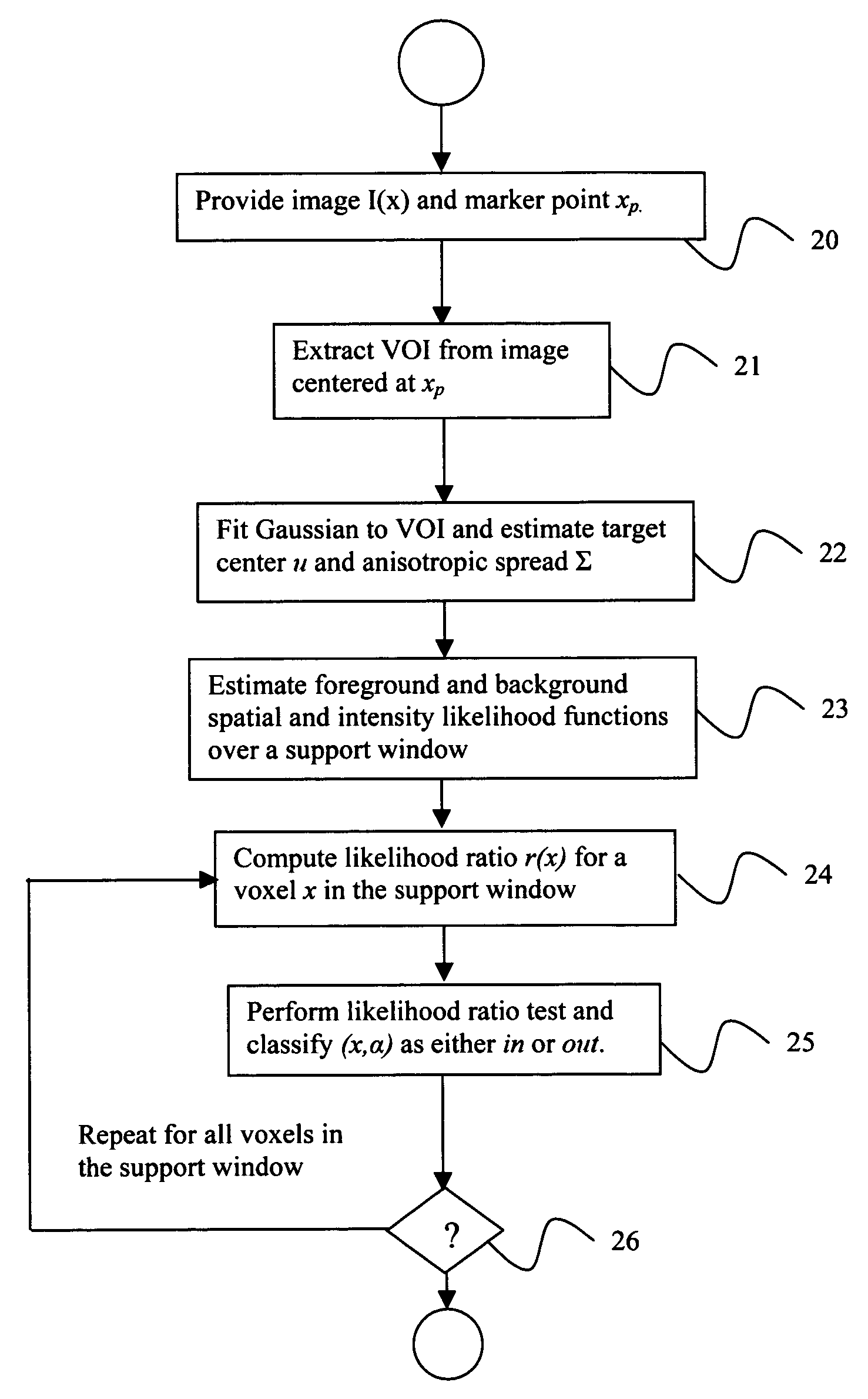
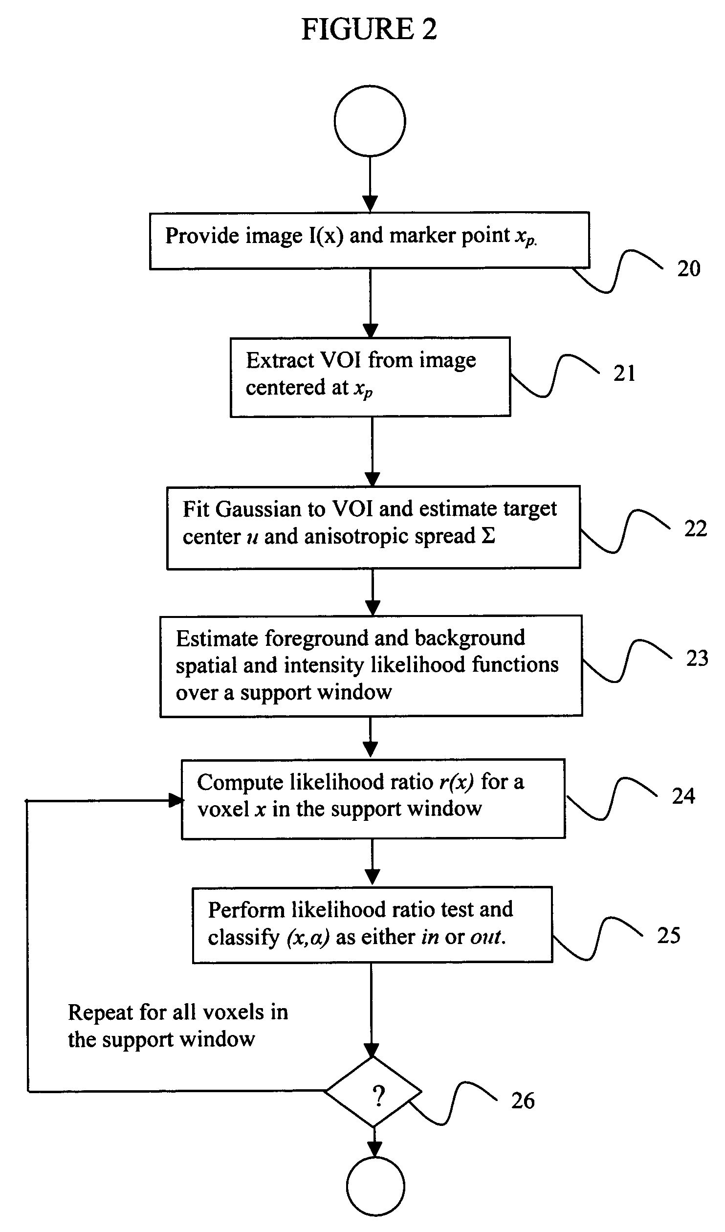
[0031]Exemplary embodiments of the invention as described herein generally include systems and methods for an efficient segmentation solution for a class of blob-like structures captured in multi-dimensional medical images. Although an exemplary embodiment of this invention is discussed in the context of segmenting a CT lung nodule, it is to be understood that the object segmentation and shape characterization methods presented herein have application to other multi-dimensional imaging modalities.

[0032]As used herein, the term “image” refers to multi-dimensional data composed of discrete image elements (e.g., pixels for 2-D images and voxels for 3-D images). The image may be, for example, a medical image of a subject collected by computer tomography, magnetic resonance imaging, ultrasound, or any other medical imaging system known to one of skill in the art. The image may also be provided from non-medical contexts, such as, for example, remote sensing systems, electron microscopy, e...


PUM


Abstract

A method for segmenting a digitized image includes providing a digitized volumetric image comprising a plurality of intensities corresponding to a domain of points in an N-dimensional space, identifying a target structure in said image, forming a window about said target structure whose size is a function of the target scale, and performing a joint space-intensity-likelihood ratio test at each point within said window to determine whether each said point is within said target structure.

Description

CROSS REFERENCE TO RELATED UNITED STATES APPLICATIONS[0001]This application claims priority from “Volumetric Tumor Segmentation using Space-Intensity Joint Likelihood Ratio Test”, U.S. Provisional Application No. 60 / 608,499 of Okada, et al., filed Sep. 9, 2004, and from “Blob Segmentation using Joint Space-Intensity Likelihood Ratio Test: Application to 3D Tumor Segmentation”, U.S. Provisional Application No. 60 / 625,027 of Okada, et al., filed Nov. 4, 2004, the contents of both of which are incorporated herein by reference.TECHNICAL FIELD[0002]This invention is directed to object segmentation in digitized medical images.DISCUSSION OF THE RELATED ART[0003]The diagnostically superior information available from data acquired from current imaging systems enables the detection of potential problems at earlier and more treatable stages. Given the vast quantity of detailed data acquirable from imaging systems, various algorithms must be developed to efficiently and accurately process image...

Claims


Application Information

Patent Timeline
Patent Type & Authority Patents(United States)
IPC IPC(8): G06K9/34
CPCG06T7/0081G06T7/0087G06T2207/30096G06T2207/20144G06T2207/10072G06T7/11G06T7/143G06T7/194
Inventor OKADA, KAZUNORIAKDEMIR, UMUTKRISHNAN, ARUN
Owner SIEMENS MEDICAL SOLUTIONS USA INC
Why Eureka
Try Eureka