Auto-stimulating cells and methods for making and using the same

a technology of auto-stimulating cells and cells, applied in the field of auto-stimulating cells and methods for making and using the same, can solve the problems of insufficient therapeutic benefit, toxicities of il-2, and inability to transfect apcs, including tumor cells, and the loss of activity of therapeutic t cells once injected into patients' bodies

Inactive Publication Date: 2008-10-14
THE TRUSTEES OF THE UNIV OF PENNSYLVANIA
View PDF7 Cites 14 Cited by
  • Summary
  • Abstract
  • Description
  • Claims
  • Application Information

AI Technical Summary

Benefits of technology

[0019]The present invention has met the above needs, by providing methods for quantitative transfer of a domain having trans-signaling and / or adhesion function onto a cell surface. Typically, the domain will be the extracellular domain having one or both of these functions. In a preferred embodiment, the extracellular domain of an immuno-regulatory molecule is used. More specifically, the present methods provide a two-step protein transfer approach, which permits delivery of graded amounts of proteins to a cell surface. The methods utilize a fusion protein comprised of at least two domains, one of which preferably encodes a molecule having immunoregulatory function. By adding the fusion protein to cells coated with a lipidated protein, fine titration of, for example, the immunoregulatory molecule's extracellular domain is achieved.

Problems solved by technology

A major limiting factor for the clinical application of therapeutic T cells is their loss of activity once injected into patients.
Costimulation is required not only for the early activation of T cells, but also for the later maintenance of their post-activation effector capacity, referred to as “effector costimulation.” In an effort to maintain therapeutic T cells in a fully activated state, activating cytokines, such as IL-2, have been administered systemically to patients receiving therapeutic T cells, albeit with IL-2 toxicities and insufficient therapeutic benefit.
For example, APCs, including tumor cells, are often poorly transfectable.
In addition, transfection proceedings are cumbersome and time-consuming.
Furthermore, expressing more than a costimulator (or coinhibitor) is difficult.
These and other issues have impeded the widespread application of gene therapy for APC and tumor cell engineering.
This method has significant limitations, including its dependence on covalent modifications that could perturb multiple proteins on cell surfaces.
No therapeutic cells in the art have been designed with membrane-embedded proteins with the capacity to trigger their own activating or inhibitory receptors.

Method used


Image

  • Auto-stimulating cells and methods for making and using the same
  • Auto-stimulating cells and methods for making and using the same
  • Auto-stimulating cells and methods for making and using the same

Examples


example 1

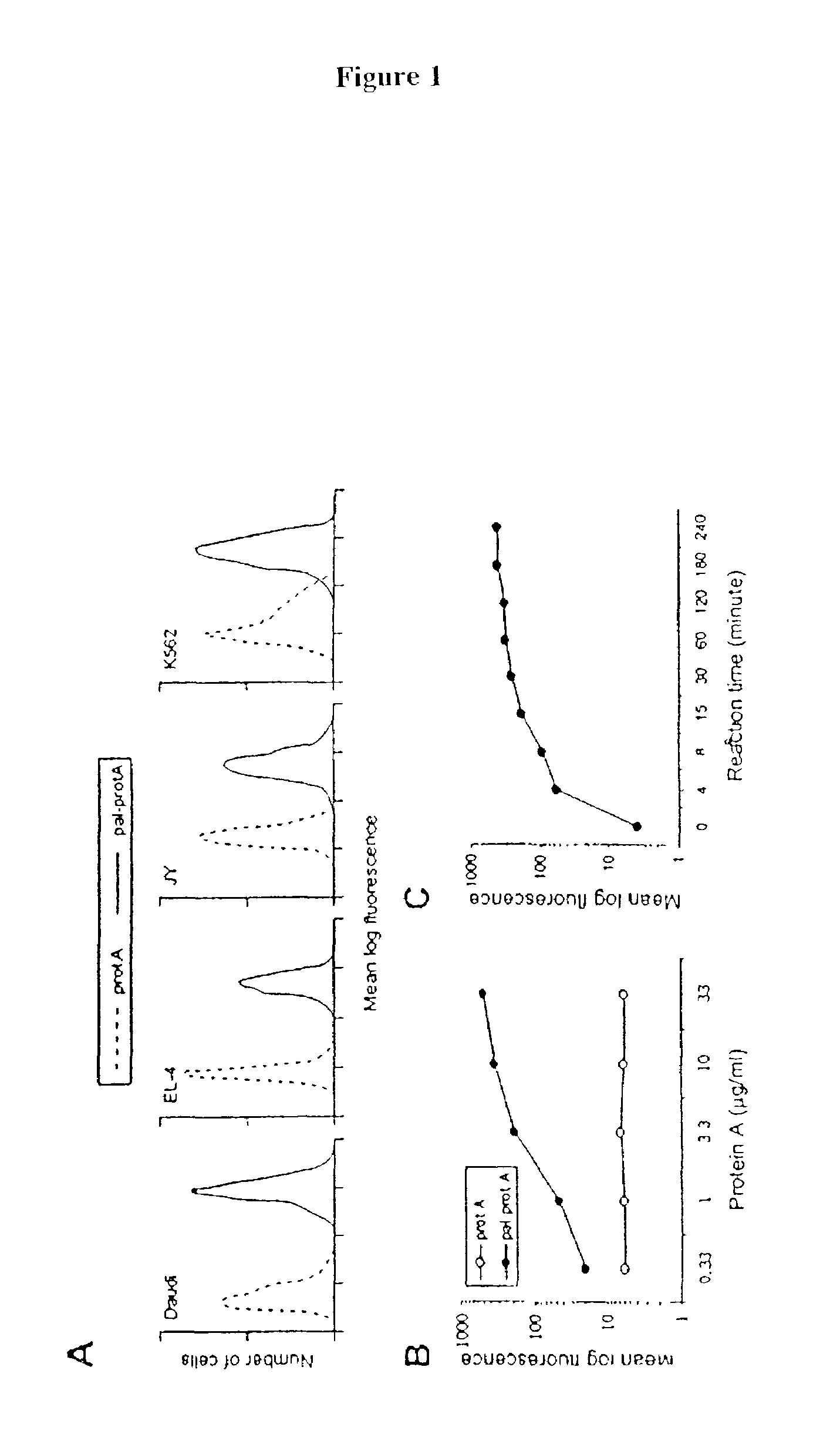
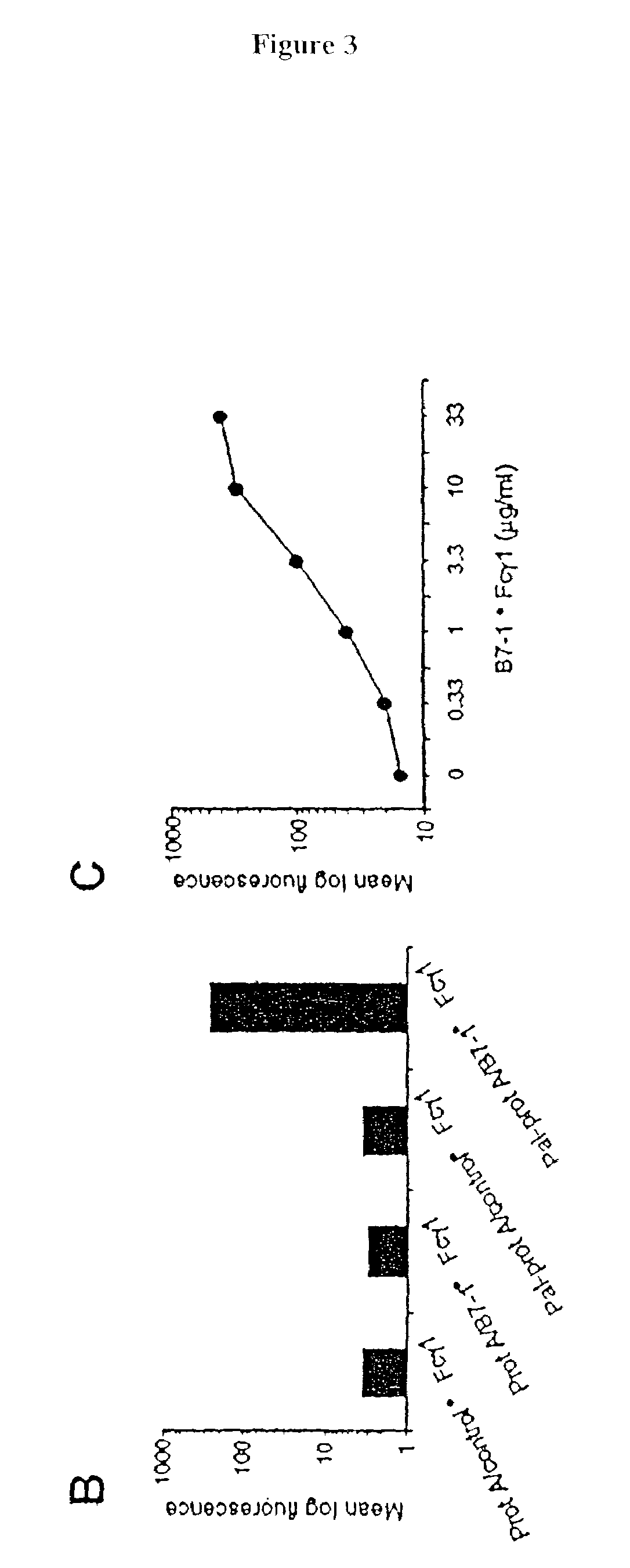
[0083]The following example demonstrates a method for transferring a B7-1•Fcγ1 fusion protein to a cell using palmitated protein A.

Palmitation of Protein A

[0084]Recombinant protein A (Calbiochem, La Jolla, Calif.) was derivatized with the N-hydroxysuccinimide ester of palmitic acid (Sigma, St. Louis, Mo.) as described by Kim and Peacock, J. Immunol Methods, 158:57 (1993). Briefly, a stock solution of the N-hydroxysuccinimide ester of palmitic acid was made, as was a solution containing protein A in a concentration of about 1.5 mg / ml. The solutions were mixed in a ratio of about 10 μg ester per ml protein and incubated at room temperature with constant mixing for about 18 h. The lipid-derivatized protein A was purified as described by Huang, et al., J. Biol. Chem., 225:8015 (1980) using a 30-ml Sephadex G-25 (Sigma) column. The protein product, referred to herein as “pal-prot A∘, was quantitated using a bicinchoninic acid kit (Bio-Rad, Richmond, Calif.), filter sterilized, and stored...

example 2

[0103]The effect of temperature on membrane-incorporated protein A was studied; the transferred protein must remain cell-bound in vivo in order to prime T-cells, which requires stable engagement of costimulators for at least several hours. It was determined that the reaction temperature at which a lipidated protein is transferred to the cell membrane has a major impact on long-term retention of the protein on the membrane. Protein transfer reactions were performed at 4° C., 25° C. or 37° C.; palmitated protein A was transferred onto K562 cells. An hB7-1•Fc was 125I-labeled and transferred to the protein A-coated cells in the manner described in Example 1. To prevent interference of endocytosis likely to occur at temperatures above 4° C., the cells were treated with the metabolic inhibitors sodium azide and 2-deoxyglucose prior to the transfer reaction.

[0104]To determine the long-term retention of the transferred protein on the cell membrane, the cells were thoroughly washed to remov...

example 3

[0105]C3H / HeN mice, purchased from Harlen (USA), Indianapolis, were immunized with a cell vaccine generated from the T-50 cell line, obtained from Avranham Hochberg, Hadassah University Hospital. The vaccine was prepared following the procedure generally outlined in Example 1, using palmitated protein A and nB7-1ωFc, m4=1 BBL•Fc, and hCD40L•Fc fusion proteins. Basically, the cells were coated with the lipidated protein A at 37° C. at a ratio of 40 μg protein A per 40×106 cells. The cells were then incubated at 4° with an equal mixture of the three fusion proteins at a ratio of 20 μg total protein per 4×10⁷ cells. The cell vaccine was injected into the mice subcutaneously at a dose of 106 cells per injection. The injections were given once a week and continued for three weeks. One week after the last injection, the animals were challenged with 106 wild-type T-50 tumor cells, injected intradermally on the rear flank. As FIG. 9 shows, the cell vaccine improved the survival rate of the ...


PUM

PropertyMeasurementUnit
concentrationaaaaaaaaaa
concentrationsaaaaaaaaaa
densityaaaaaaaaaa

Abstract

Methods for transferring one or more proteins to a cell are disclosed. The protein or proteins to be transferred are in the form of a fusion protein, and contain at least one domain encoding for a protein or peptide having trans signaling and / or adhesion function. The fusion protein is transferred to a cell by binding to a lipidated protein, which has been incorporated into the cell membrane. In an additional aspect of the invention, methods of making fusion proteins having cis signaling capabilities, as well as the ability to bind with receptors on the cell's own surface, are provided. Fusion proteins incorporating GPI or a homing element, and a costimulator or inhibitor domain can also be directly transferred to the cell surface. Methods for using cells which have undergone protein transfer according to the present methods are also disclosed. This includes use in a cancer vaccine, use for treatment of cancer or autoimmune disease, and use in determining costimulator threshold levels.

Description

CROSS-REFERENCE TO RELATED APPLICATION[0001]This application claims priority under 35 USC § 119(e) to Provisional Application Ser. No. 60 / 398,050, filed Jul. 22, 2002, and is a continuation-in-part of application Ser. No. 09 / 957,056. filed Sep. 20, 2001 , which is a divisional application of09 / 476,828, filed Jan. 3, 2000, now U.S. Pat. No. 6,316,256, expressly incorporated herein by reference.[0002]This work was supported in part by Grants R01 CA-74958 and R01 AI-31044 from the National Institutes of Health.FIELD OF THE INVENTION[0003]The present invention relates to novel methods for transferring one or more proteins to a cell. In addition to other applications, the methodology is useful in the treatment of cancer and autoimmune diseases, and for determining costimulator activation thresholds and cooperative interactions among costimulators. The invention further relates to cells painted with extrinsic proteins that are capable of triggering receptors on the surfaces of the same ce...

Claims


Application Information

Patent Timeline
Patent Type & Authority Patents(United States)
IPC IPC(8): C12N5/00C12N5/02C12N5/06C12N5/16A61K35/12C12N15/09A61K35/14A61K35/17A61K35/28A61K35/30A61K35/32A61K35/34A61K35/36A61K39/00A61K47/48A61P31/12A61P35/00A61P37/06C07K14/705C12N5/08C12N5/10
CPCA61K39/0011A61K47/48246C07K14/70525C07K14/70532C07K14/70575C12N5/0006A61K35/17A61K39/00A61K2039/5158A61K2039/55516A61K2039/6031C07K2319/00C07K2319/20C07K2319/30C12N2501/51C12N2501/58A61K38/00A61K47/64A61P31/12A61P35/00A61P37/06
Inventor TYKOCINSKI, MARK L.ZHENG, GUOXING
Owner THE TRUSTEES OF THE UNIV OF PENNSYLVANIA
Why Eureka
Try Eureka