Method for diagnosing increased risk of preeclampsia
a preeclampsia and increased risk technology, applied in the field of diagnosis and treatment of conditions, can solve the problems of preeclampsia and short invasion, and achieve the effect of reducing the invasion of trophoblasts
- Summary
- Abstract
- Description
- Claims
- Application Information
AI Technical Summary
Benefits of technology
Problems solved by technology
Method used
Image
Examples
example i
Materials and Methods
Establishment of Human Trophoblast Villous Explant Culture
[0097]Villous explant cultures were established from first trimester human placentae by a modification of the method of Genbacev et al. (21). First trimester human placentae (5-8 weeks gestation) were obtained from elective terminations of pregnancies by dilatation and curettage. Placental tissue was placed in ice-cold phosphate bufferd saline (PBS) and processed within two hours of collection. The tissue was washed in sterile PBS, and aseptically dissected using a incroscope to remove endometrial tissue and fetal membranes. Small fragments of placental villi (15-20 mg wet weight) were teased apart and placed on a transpant Biopore membrane of 12-mm diameter Millicell-CM culture dish inserts with a pore size of 0.4 μm (Millipore Corp. Bedford, Mass.). The inserts were precoated with 0.2 ml of undiluted Matrigel (Collaborative Research Inc), polymerized at 37° C. for 30 min. and transferred in a 24 well cu...
example 2
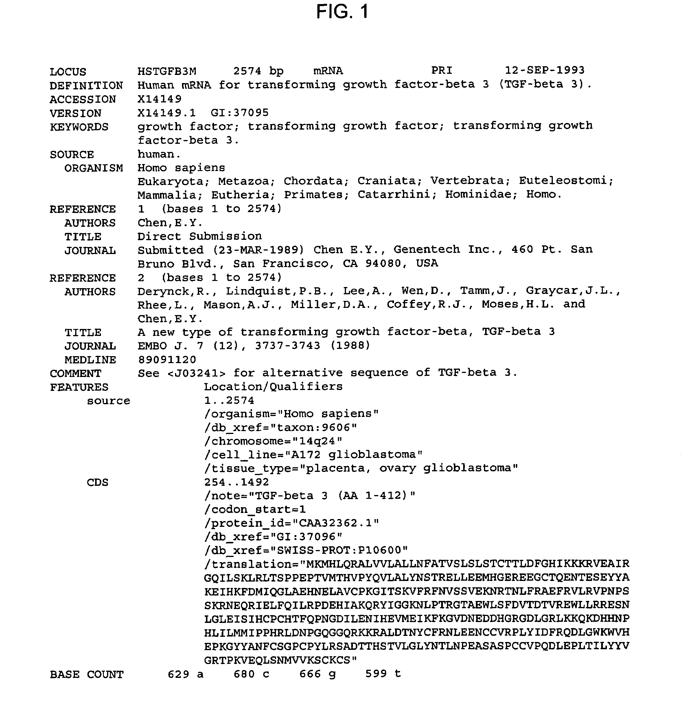
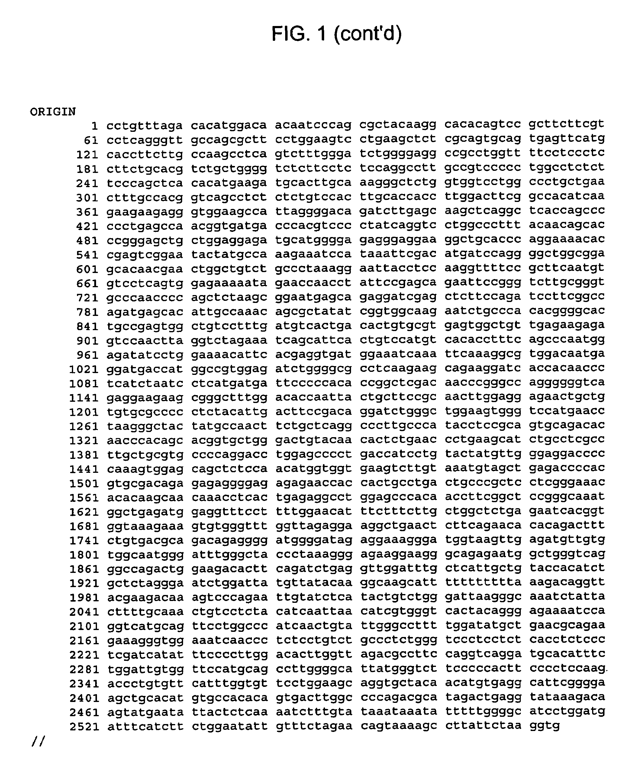
[0119]The present experiments were conducted to define the precise components that endogenously regulate trophoblast invasion. Using human villous explants of 5-7 weeks gestation it was observed that while trophoblast cells remain viable they do not spontaneously invade into the surrounding matrigel. In contrast, trophoblast cells from 9-13 weeks explants spontaneously invade the matrigel in association with an upregulation of fibronectin synthesis and integrin switching. Trophoblast invasion at 5-7 weeks can be induced by incubation with antisense to TGF-β3, TGFβ receptor I (ALK-1) or TGFβ receptor II. Only minimal invasion occurred in response to antisense to TGFβ1 and antisense TGFβ2 failed to induce invasion. These data suggest that TGF-β3 via the ALK-1-receptor II complex is a major regulator of trophoblast invasion in vitro. To determine whether this system may also operate in vivo, immunohistochemical staining was conducted for TGF-β1 and -3 and for TGFβ receptor I and II in ...
example 3
[0120]Studies were carried out to determine if shallow trophoblast invasion in preeclampsia was associated with an abnormally sustained inhibition of invasion by TGF-β. In particular, the expression / distribution of the different TGF-β isoforms and their receptors was investigated using immunohistochemical analysis in normal placentae at 7-9 weeks (at the onset of trophoblast invasion) at 12-13 weeks (the period of peak invasion), in control placentae between 29 and 34 weeks and in preeclamptic placentae ranging from 27 to 34 weeks. In normal placentae. TGF-β3 expression was markedly reduced with advancing gestational age. Expression was high in cyto- and syncytiotrophoblast cells at 7-9 weeks of gestation but was absent in villous tissue at 12-13 weeks and at 29-34 weeks of gestation. A similar decline in positive immunoreactivity against TGF-β receptor I and II was also observed over this time period. In contrast, in preeclamptic placentae between 27-34 weeks of gestation, strong s...
PUM
| Property | Measurement | Unit |
|---|---|---|
| time | aaaaa | aaaaa |
| time | aaaaa | aaaaa |
| pore size | aaaaa | aaaaa |
Abstract
Description
Claims
Application Information
 - R&D
- Intellectual Property
- Life Sciences
- Materials
- Tech Scout
- Unparalleled Data Quality
- Higher Quality Content
- 60% Fewer Hallucinations
Browse by: Latest US Patents, China's latest patents, Technical Efficacy Thesaurus, Application Domain, Technology Topic, Popular Technical Reports.
© 2025 PatSnap. All rights reserved.Legal|Privacy policy|Modern Slavery Act Transparency Statement|Sitemap|About US| Contact US: help@patsnap.com


