Spectral imaging of biofilms

a biofilm and raman spectroscopy technology, applied in the field of raman spectroscopy identification of biofilms, can solve the problem that biofilms are more difficult to get rid, and achieve the effect of reducing the number of biofilms

Active Publication Date: 2008-11-11
CHEMIMAGE CORP
View PDF10 Cites 5 Cited by
  • Summary
  • Abstract
  • Description
  • Claims
  • Application Information

AI Technical Summary

Benefits of technology

[0031]For instance, the methods and devices described herein can be used to identify the presence or absence of sessile forms of a microorganism, such as in settings in which it is important to detect biofilm formation at an early time—before heavy accumulation of biofilm material can occur. The methods and devices described herein can also be used to determine the effect of a composition (e.g., an antibiotic) on cells of a biofilm. Such analysis facilitates development of effective compositions for countering accumulation and survival of biofilms.
[0044]In a preferred embodiment, a Raman scattering property of the microorganism is examined. Use of Raman-shifted scattered light decreases interference with light that is reflected or elastically scattered by the sample, permitting more detailed analysis of the sample than might otherwise be possible. In order to assess Raman-shifted scattered light, a monochromatic light source should be used to illuminate the sample and a filter that substantially prevents transmission of light having the same wavelength as the light source should be interposed between the sample and the Raman detector. Raman spectroscopy is a preferred method because the Raman spectrum of a sample can be used to identify a wide variety of materials and organisms, each of which can exhibit characteristic Raman spectral properties that facilitate their identification.
[0049]Whether a microorganism occurs in a planktonic or sessile form can have important implications, especially in medical contexts. Many parts of the body of humans and other animals are normally aseptic. Occurrence of a microorganism in such parts (in any form) is usually a sign of disease. At other parts of the body (e.g., skin and vaginal surfaces and much of the lining of the digestive tract), a variety of microorganisms normally occur when the animal is in a healthy state. Ability to distinguish planktonic and sessile forms of a microorganism at a body location can enable one to distinguish pathologic and non-pathologic states in a tissue sample. Even in normally sterile tissues, the planktonic / sessile forms of a pathogenic microorganism can influence the most desirable form of medical intervention. The methods and devices described herein can be used for such purposes by facilitating both identification of microorganisms that occur at a body site and the planktonic / sessile form of the organisms detected there.
[0070]The image in FIG. 5A was taken using the Falcon (RTM) device at 100× magnification, at wavenumbers from 800 to 3100 cm−1 at a laser power of 190 milliwatts. Three regions of interest 510, 520, and 530 are highlighted within the field of view shown in FIG. 5A. The two mucosal colony areas 510 and 530 and the aluminum background area 520 exhibit different spectral profiles. FIGS. 5B and 5C show the corresponding Raman spectra 530b and 530c for region of interest 530, Raman spectra 520b and 520c for region of interest 520 and Raman spectra 530b and 530c for region of interest 530. The information from the Raman Chemical Image was overlaid upon the bright field reflectance video capture image taken very close to the same field of view. The information from this overlay (shown in FIG. 6) confirmed that the chemical information signal originated from the mucosal colonies and included minimal interference from the aluminum background.

Problems solved by technology

Biofilms tend to be more difficult to get rid of than individual microorganisms or non-film colonies of microorganisms.

Method used


Image

  • Spectral imaging of biofilms
  • Spectral imaging of biofilms
  • Spectral imaging of biofilms

Examples


examples

[0062]The subject matter is now described with reference to the following Examples. These Examples are provided for the purpose of illustration only, and the subject matter is not limited to these Examples, but rather encompasses all variations which are evident as a result of the teaching provided herein.

Imaging of Planktonic and Sessile Pseudomonas aeruginosa Cells

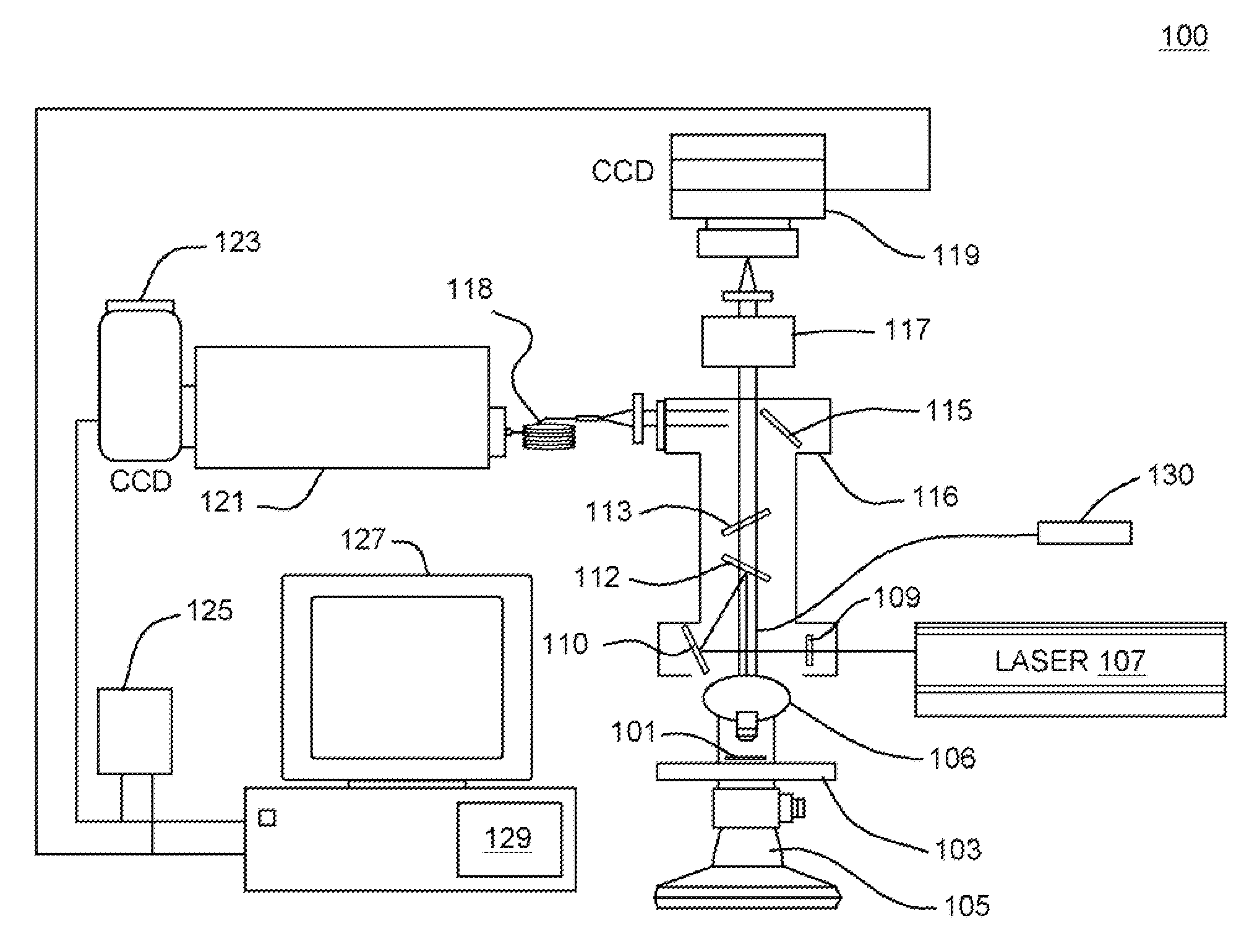
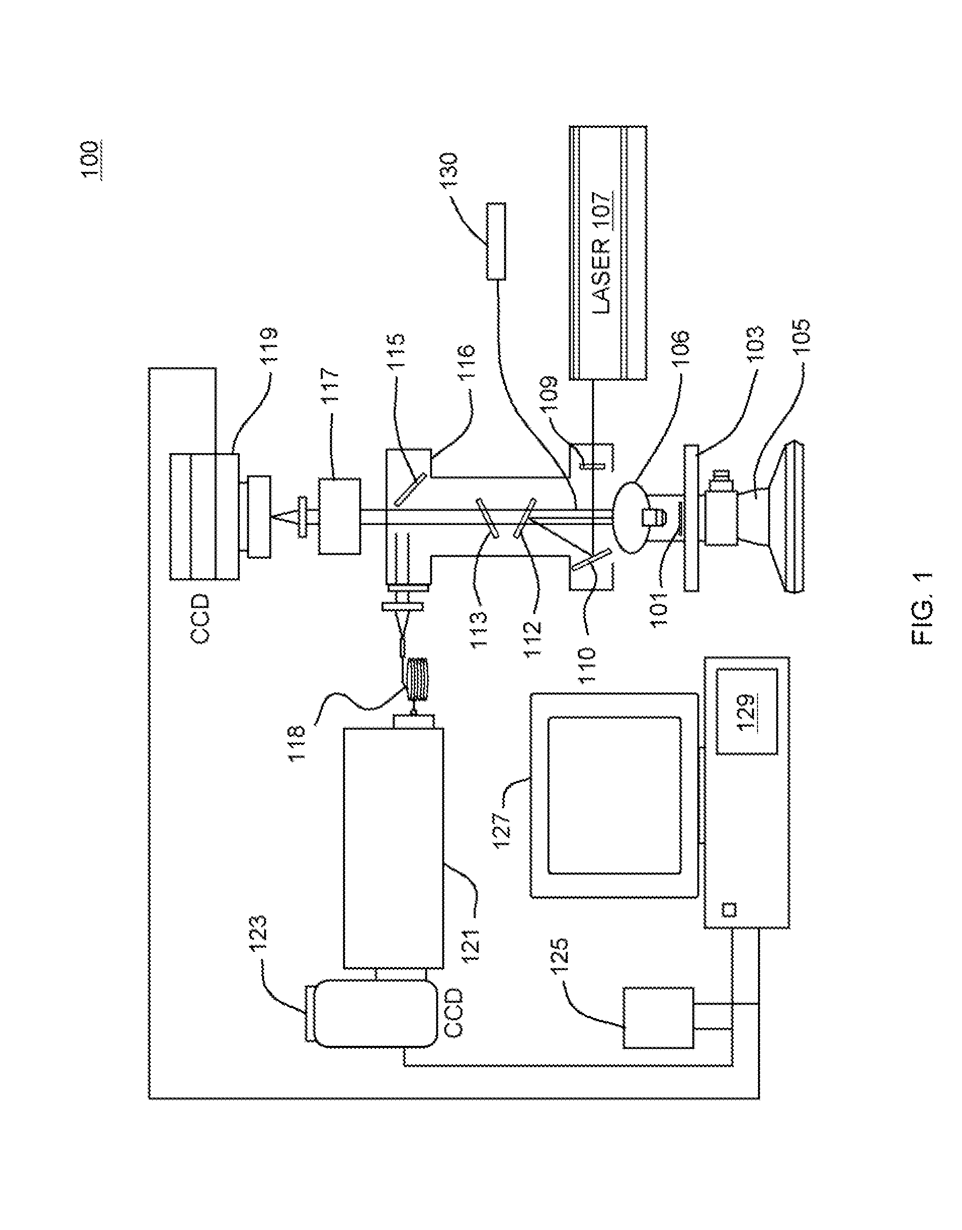
[0063]The experiments described in this Example involved imaging of the planktonic and sessile (biofilm) forms of Pseudomonas aeruginosa. The planktonic and sessile organisms were deposited on aluminum-coated slides and prepared the samples for imaging.

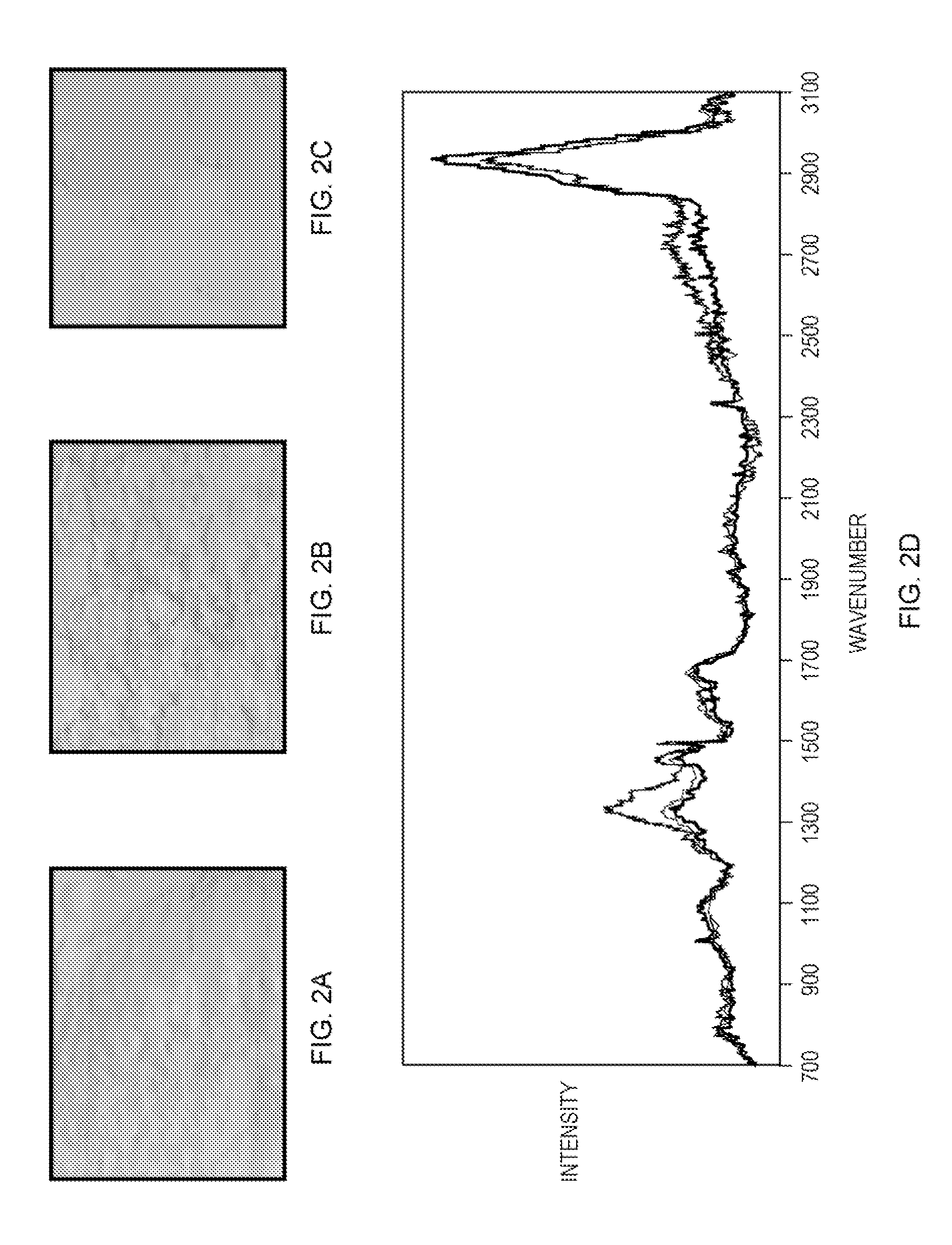
[0064]Planktonic cultures were filtered and then printed to a coupon directly and in dilutions of 1:10 and 1:100 (corresponding to FIGS. 2A, 2B, and 2C, respectively). Biofilm cultures were directly grown on slides and air-dried. The samples were characterized by dispersive Raman spectroscopy. Multiple spectra were taken of all forms and dilutions. After enhancing the spec...


PUM


Abstract

A spectroscopic method and system to identify a biofilm of a microorganism. A sample containing a sample microorganism is irradiated with substantially monochromatic radiation. A Raman data set is obtained based on radiation scattered from the irradiated sample. A database is searched in accordance with the Raman data set in order to identify a known Raman data set from the database. The database contains a plurality of known Raman data sets where each known Raman data set is associated with a known sessile form of a corresponding known microorganism. A sessile form of the sample microorganism is identified based on the known Raman data set identified by the searching.

Description

RELATED APPLICATIONS[0001]This application claims the benefit of U.S. Provisional Appl. No. 60 / 734,839, filed Nov. 9, 2005, entitled “Spectral Imaging of Biofilm” which is incorporated herein by reference in its entirety.FIELD OF THE DISCLOSURE[0002]This application generally relates to the identification of biofilms by Raman spectroscopy.BACKGROUND OF THE DISCLOSURE[0003]Microorganisms sometimes grow in films or mats, in which not all microorganisms in the film are identical. The form, function, composition, and metabolic state of cells can vary depending on their location in the film. Cells on the exterior surface of the film can exhibit different properties and functions than cells in the interior of the film or cells at the surface of the film that is in contact with a substrate.[0004]Biofilms are usually composed of both an extracellular matrix and cells. The matrix can anchor the cells to the substrate and provide a matrix in which the cells can live. Common biofilm materials ...

Claims


Application Information

Patent Timeline
Patent Type & Authority Patents(United States)
IPC IPC(8): G01J3/44
CPCG01J3/02G01J3/0227G01J3/0291G01J3/44G01N21/65G01N21/658G01N2021/656G01N2201/129
Inventor MAIER, JOHNCOHEN, JEFFREY
Owner CHEMIMAGE CORP
Why Eureka
Try Eureka