Method and arrangement relating to x-ray imaging

a technology of automatic exposure control and x-ray imaging, which is applied in the field of method and arrangement of automatic exposure control in an x-ray apparatus, to achieve the effect of improving the exposure over the breast and low attenuation of x-rays

Active Publication Date: 2009-02-24
PHILIPS DIGITAL MAMMOGRAPHY SWEDEN
View PDF8 Cites 20 Cited by
  • Summary
  • Abstract
  • Description
  • Claims
  • Application Information

AI Technical Summary

Benefits of technology

[0015]A problem with state-of-the art mammography systems and corresponding AECs is that the exposure is not optimised in every area of the image, the systems can only deliver and average exposure that will be too high in thin areas of the breast and in fatty areas of the breast with low attenuation of the x-rays while it will be to high in dense areas of the breast with predominantly glandular tissue that is more opaque to x-rays. The current invention solves this problem since the pre-exposure is combined with the diagnostic exposure in a real-time system and the exposure needs only to be averaged in one dimension (orthogonal to the scan direction) while it may be fully optimised in the other dimension (the scan direction). Thus a significantly improved exposure over the breast can be achieved.
[0016]Problems related to scanning systems are to obtain precise and stable mechanical motion over time and wear of the mechanical parts. If the position of the scanning detector is accurately measured under all conditions, something that is relatively easy to achieve, and the system is read out at triggers from the measurement of the position, e.g. every 50 μm, and the time between triggers is measured accurately with an external clock, the measured exposure at the detector can be normalized in such a way that the system becomes insensitive to mechanical instability; thus, a very robust system is achieved. Input to AEC will be the normalised signal from a detector, i.e. the signal per true 50 μm, the time the exposure was collected measured with the external clock. The time will be used to calculate actual velocity 50 μm / t and also non-normalise the detector signal.
[0018]The proposed method scans the object (breast) with the detector that controls the exposure time. Conventional systems have the same exposure time for each point of the breast. The method according to the invention controls the exposure of each point based on the signal or density in the very same point, at least in the scan direction. Conventional film based systems control the exposure time by terminating the exposure. The proposed system may control the exposure in real time during the acquisition by altering any of scan speed, tube current (mA), tube voltage (kV), anode material and filter. According to the preferred embodiment of the invention, the system controls the exposure time by varying the scan-speed and thus the exposure time of each point in the scanning direction. The scanning speed is based on the exposure substantially in the every point being exposed.
[0019]Thus, the object of the present invention according to preferred embodiments of the invention is to provide a novel method and arrangement for Automatic Exposure Control, which lacks the drawbacks of the prior art. The invention beside conventional x-ray imaging systems is also suitable for X-ray diagnostic apparatus using photon-counting technique. This invention is particularly suitable for photon counting detectors, since there is a simple relationship between counted photons and SNR. Also the detector is very linear which further means a simple relationship to scan speed and target signal. Preferably, the object of the present invention is to provide a system based on a non-constant scan-velocity, which is controlled in substantially real-time during the scan.
[0023]Most preferably, the feedback is substantially real-time and controls the scan speed of the detector.
[0024]In one embodiment, the exposure of each point along an x-axis is controlled based on the count rate of the said region and thus the entire image has a controllable signal level along the first dimension at least in said region in the second dimension. The method further comprises minimizing total scan time by areas not covered by dense objects being scanned faster and thus exposed shorter. Preferably, the detector itself is used to control the exposure.

Problems solved by technology

A problem with state-of-the art mammography systems and corresponding AECs is that the exposure is not optimised in every area of the image, the systems can only deliver and average exposure that will be too high in thin areas of the breast and in fatty areas of the breast with low attenuation of the x-rays while it will be to high in dense areas of the breast with predominantly glandular tissue that is more opaque to x-rays.
Problems related to scanning systems are to obtain precise and stable mechanical motion over time and wear of the mechanical parts.

Method used


Image

  • Method and arrangement relating to x-ray imaging
  • Method and arrangement relating to x-ray imaging
  • Method and arrangement relating to x-ray imaging

Examples


Embodiment Construction

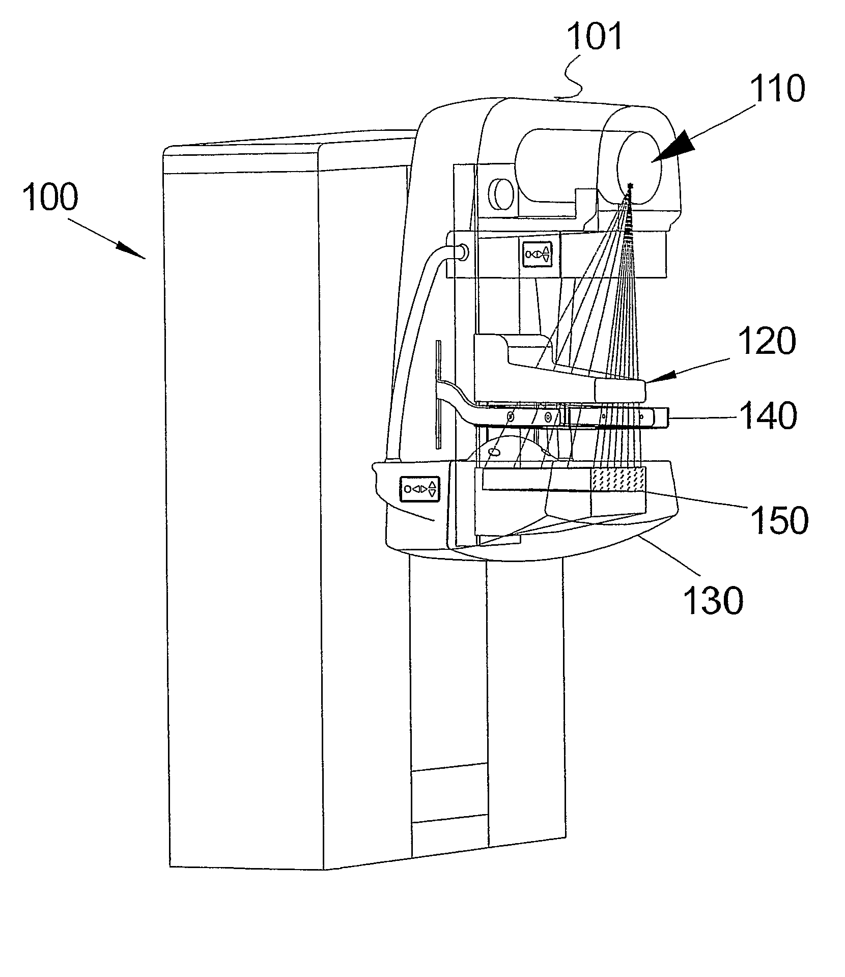
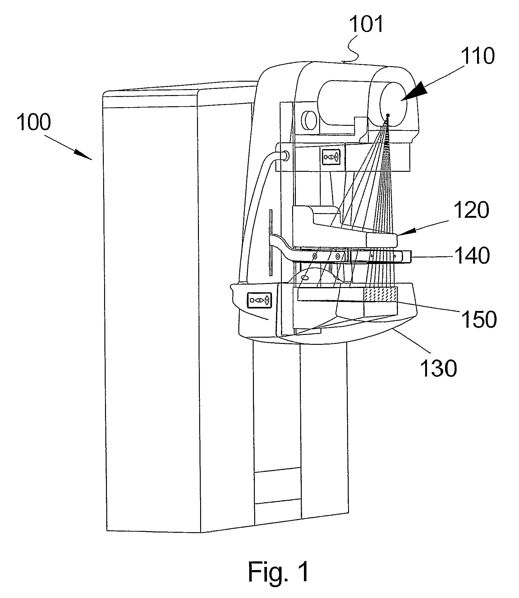
[0043]FIG. 1 illustrates an X-ray imaging system 100, according to one preferred embodiment, based on a photon-counting detector that scans the image field in one dimension that is referred to as the x-dimension. The system 100 comprises an X-ray source (tube) 110 arranged in a housing 101, patient support 130 and pre-collimator housing 120 and compression paddle. A collimator 140 is arranged in a collimator support structure and the patient support 130 includes an array of detectors 150.

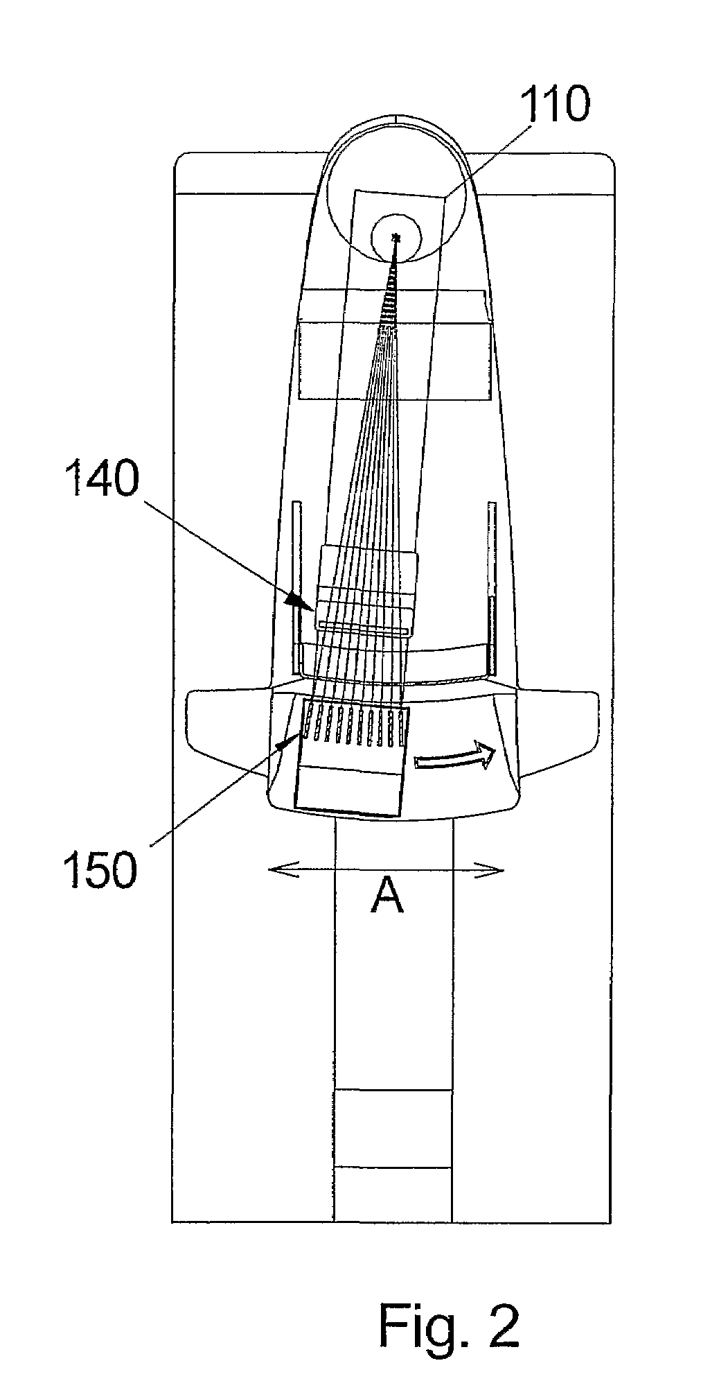
[0044]As it is illustrated in FIG. 2, the X-ray source 110 and detector array 150 are arranged to be displaced radially with the source 110 in the centre, thus scanning the section A. An image is acquired by scanning a detector across the image field. Whenever the detector has scanned a predefined distance, the number of photon counts collected is read-out and the counter is reset (zeroed). This means that the distance the detector moves between readouts defines the pixels in the scan direction. In ...


PUM


Abstract

The present invention relates to a method and arrangement for controlling exposure in an e-ray apparatus, for depicting an object. The apparatus comprises an x-ray source and a displaceable detector being arranged to move with a controllable speed across an image exposure area. The method comprises the step of: acquiring a signal relating to photons incident on at least a part of the detector, comparing said acquired signal with a target value, and controlling the speed of detector, displacement with respect to the result of the comparison.

Description

CROSS REFERENCE TO RELATED APPLICATIONS[0001]This application claims priority benefit under 35 U.S.C. §365(a) of International Patent Application No. PCT / SE05 / 000194 filed 14 Feb. 2005, which, in turn, claims priority benefit of Swedish Patent Application Serial No. 0400347-1 filed Feb. 13, 2004. The entire contents of both applications are hereby incorporated in total by reference.THE FIELD OF THE INVENTION[0002]The present invention relates to a method and arrangement for Automatic Exposure Control (AEC) in an X-ray apparatus.THE BACKGROUND OF THE INVENTION[0003]AEC is a mode of operation of an X-ray machine by which the x-ray output is automatically controlled and terminated when a pre-set radiation exposure to the image receptor is reached. It may also operate in such a way that a pre-exposure is made before the diagnostic exposure and a sensor, e.g. part of the sensor used for the diagnostic imaging, is measuring the outcome of the pre-exposure and from the measured data the op...

Claims


Application Information

Patent Timeline
Patent Type & Authority Patents(United States)
IPC IPC(8): H05G1/38A61B6/00F16B1/00F16B5/00H05G
CPCA61B6/502A61B6/542F16B5/004F16B2200/79H05G1/38
Inventor ASLUND, MAGNUS
Owner PHILIPS DIGITAL MAMMOGRAPHY SWEDEN
Why Eureka
Try Eureka