
Automated radiograph classification using anatomy information

a radiograph and anatomy information technology, applied in the field of automatic classification of digital radiographs, can solve the problems of difficult automatic and efficient processing of computers, affecting the efficiency of routine medical practice and patient care, and affecting the quality of radiographs, so as to achieve the effect of robust classification

Active Publication Date: 2009-12-01
CARESTREAM HEALTH INC
View PDF9 Cites 13 Cited by
  • Summary
  • Abstract
  • Description
  • Claims
  • Application Information

AI Technical Summary

Benefits of technology

[0016]An advantage of the present invention is that the method provides an efficient way to extract the features of anatomy in images and a deliberate design of scale, rotation and translation invariant shape classifier, which makes the classification more robust.
is that the method provides an efficient way to extract the features of anatomy in images and a deliberate design of scale, rotation and translation invariant shape classifier, which makes the classification more robust.

Problems solved by technology

His provides flexibility to users, but also introduces the difficulty in setting an appropriate tone scale for image display. n optimal tone scale, in general, is dependent upon the examination type, the exposure conditions, the image acquisition device and the choice of output devices as well as the preferences of the radiologist. among them, the examination type is one determinant factor, since it is directly related to the characteristics of signal and clinical important parts in images.
This step is often skipped or the incorrect information is recorded in the image header, which hinders the efficient use of images in routine medical practice and patient care.
However, it is difficult to let a computer automatically and efficiently analyze contents in images and classify images, since image data is more structurally complex than other kinds of data and the way by which human beings capture the image contents, group image features into meaningful objects and attach semantic descriptions to images through model matching has not been fully understood to automate the analysis procedure.
Furthermore, segmenting an image into regions corresponding to individual objects, extracting features from the image that capture the perceptual and semantic meanings, and matching the image with the proposed model based on extracted features also make the analysis problem more challenging.
Such clustering-based classification schemes are usually time-consuming and of limited practical use since little of the image object semantics is explicitly modeled.
Another problem is that these systems use images collected from the world wide web.
Unfortunately, the color-based features are not available in most medical images.
However, the manual generation of the template images is quite time consuming, and more crucial, it is highly observer dependent, which may introduce error in the classification. J. Dahmen, et al (“Classification of Radiographs in the ‘Image Retrieval in Medical Application’-System”, Procs 6th International RIAO Conference on Content-Based Multimedia Information Access, Paris, France, 2000; 551-566) teach a method to classify radiographs by using a new distortion model and an extended version of simard's tangent distance with a kernel density based classifier.
Both of the above methods suffer problems in handling rotation and translation variance of anatomy in radiographs.
So the result measures cannot accurately represent the features in radiographs.
In addition, no preprocessing is implemented in the above methods.
For example, the unexposed regions caused by the blocking of the x-ray collimator during the exposure may result in a significant white borders surrounding the image.
If such regions are not removed in a pre-processing step and therefore used in the computation of similarity measures, the classification results can be seriously biased.
However, the extracted features are not sufficient to fully represent the characteristics of the edge direction histogram.
As a result, the classifier's performance is hard to be improved.

Method used


Image

  • Automated radiograph classification using anatomy information
  • Automated radiograph classification using anatomy information
  • Automated radiograph classification using anatomy information

Examples


Embodiment Construction

[0026]The following is a detailed description of the preferred embodiments of the invention, reference being made to the drawings in which the same reference numerals identify the same elements of structure in each of the several figures.

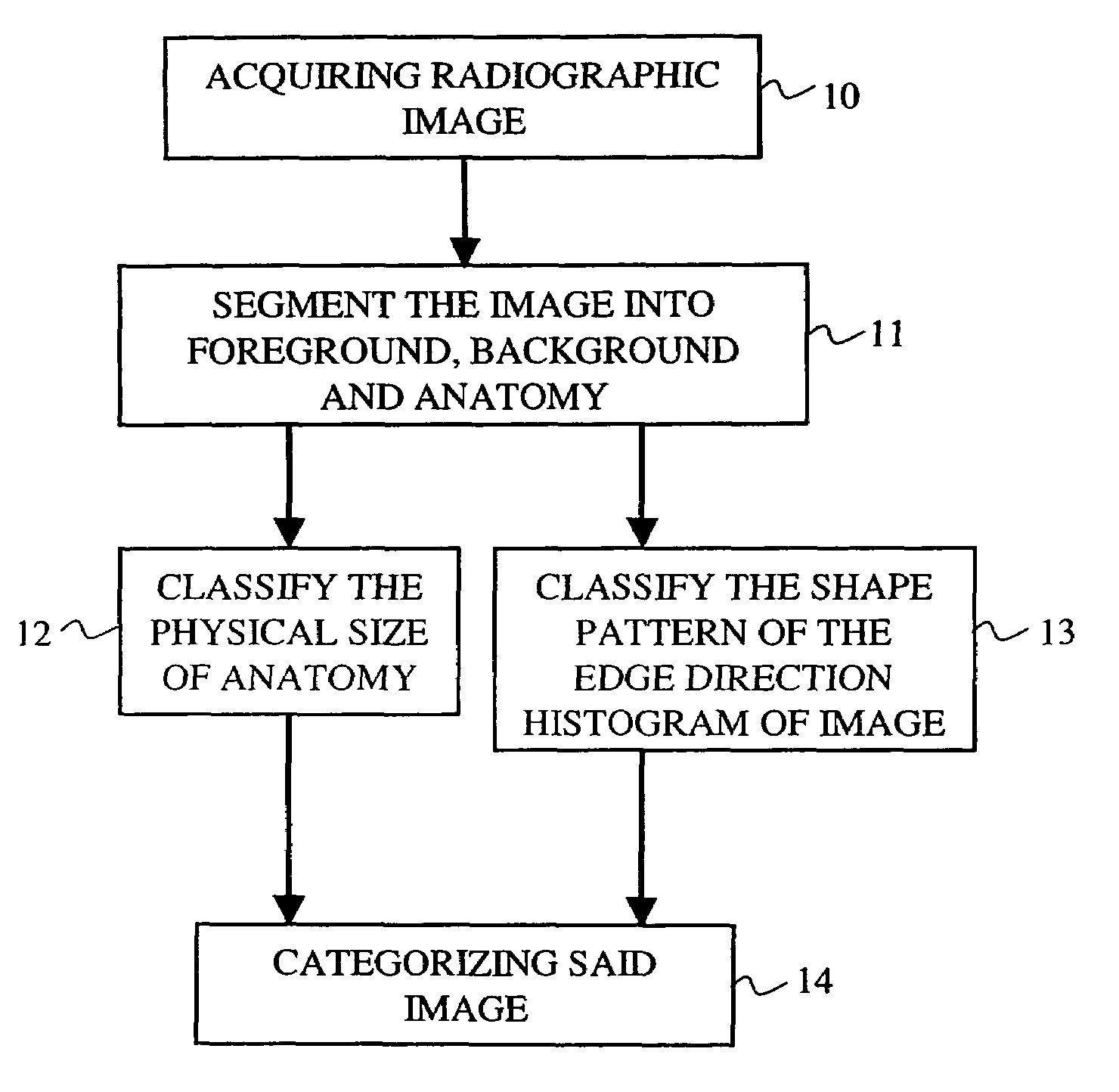
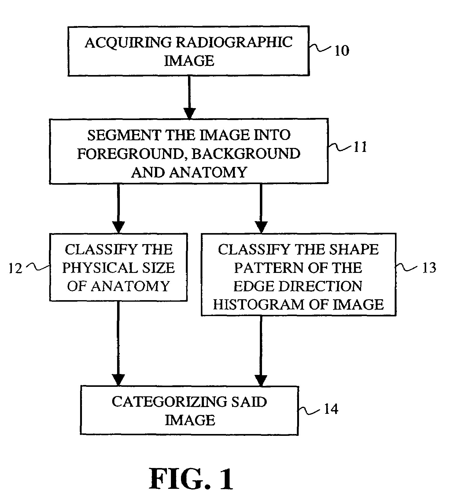
[0027]The present invention discloses a method for automatically classifying radiographs. A flow chart of a method in accordance with the present invention is generally shown in FIG. 1. As shown in FIG. 1, the method includes three stages. In the first stage, the input radiograph (box 10) is segmented (step 11) into three regions, which are: a collimation region (foreground), a direct exposure region (background) and a diagnostically relevant region (anatomy). Then, two classifications are performed on the image: one is based on a size of the anatomy (box 12), the other is based on a shape of the anatomy (box 13). Afterwhich, the results from these two classifications are combined and the input image is categorized into one of classed, for example, ...


PUM


Abstract

A method for automatically classifying a radiograph. A digital radiographic image is acquired, wherein the image is comprised of a matrix of rows and columns of pixels. The digital image is segmented into foreground, background, and anatomy regions. A physical size of the anatomy region is classified. An edge direction histogram of the anatomy region is generated and a shape pattern of the edge direction histogram is classified. Based on the physical size classification and the shape pattern classification, the image is categorized.

Description

CROSS REFERENCE TO RELATED APPLICATIONS[0001]Reference is made to, and priority is claimed from, U.S. Provisional Application No. 60 / 630,286 entitled “AUTOMATED RADIOGRAPH CLASSIFICATION USING ANATOMY INFORMATION”, filed on Nov. 23, 2004 in the names of Luo et al, and which is assigned to the assignee of this application, and incorporated herein by reference.[0002]Reference is made to commonly assigned application U.S. Ser. No. 10 / 993,055 entitled “DETECTION AND CORRECTION METHOD FOR RADIOGRAPH ORIENTATION”, filed on Nov. 19, 2004 in the names of Luo et al, and which is assigned to the assignee of this application, and incorporated herein by reference.[0003]Reference is further made to U.S. Provisional Application No. 60 / 630,270, entitled “METHOD FOR AUTOMATIC SHAPE CLASSIFICATION”, filed on Nov. 23, 2004 in the names of Luo et al, and which is assigned to the assignee of this application, and incorporated herein by reference.[0004]Reference is further made to U.S. Provisional Appli...

Claims


Application Information

Patent Timeline
Patent Type & Authority Patents(United States)
IPC IPC(8): G06K9/00A61B5/05G06V10/50
CPCG06F17/30259G06K9/4642G06K9/4647G06T7/0083G06T7/0012G06T2207/30004G06T2207/10116G06T2207/20144Y10S128/922G06T7/12G06T7/194G06F16/5854G06V10/507G06V10/50
Inventor LUO, HUIWANG, XIAOHUI
Owner CARESTREAM HEALTH INC
Learn More