Forms of prostate specific antigens and methods for their detection

a prostate specific and antigen technology, applied in the field of forms, can solve the problems of difficult development of ppsa-specific mabs, no suitable mabs,

Inactive Publication Date: 2010-02-09
HYBRITECH
View PDF6 Cites 2 Cited by
  • Summary
  • Abstract
  • Description
  • Claims
  • Application Information

AI Technical Summary

Benefits of technology

[0010]The present invention also encompasses a method for detecting proPSA in a human tissue or physiological fluid. This aspect of the invention is based on the discovery that proPSA exists stably in biological fluid as part of free PSA and may serve as a useful marker for prostate cancer. Specifically, several inactive precursor forms of PSA, such as, but not limited to [−2], [−4] and [−7]pPSA forms, have been identified and detected in serum. In accordance with the present invention, the identified precursor forms of PSA do not form a complex with ACT and exist as stable and free PSA in serum. The measurement of these inactive precursor forms of PSA may provide important information regarding the detection, monitoring and staging of prostate cancer.

Problems solved by technology

Because of the minor structural differences between pPSA and PSA, the development of pPSA-specific mAbs has been extremely difficult in the past.
In the literature, efforts specifically designed to generate pPSA mAbs have yielded no suitable mAbs12.

Method used


Image

  • Forms of prostate specific antigens and methods for their detection
  • Forms of prostate specific antigens and methods for their detection
  • Forms of prostate specific antigens and methods for their detection

Examples


example 1

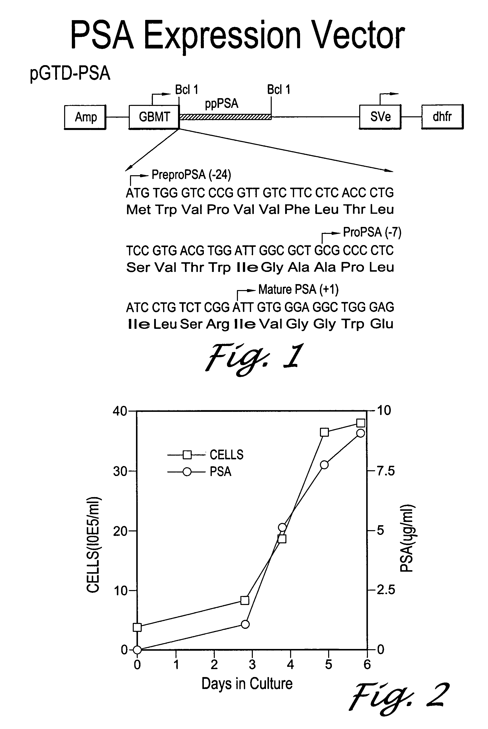
Expression of pPSA in Mammalian Cells

[0105]The cDNA for PSA was cloned into the pGT-d vector under the control of the GBMT promoter using an approach similar to the one described for hK2 by Kumar et al. To study the expression of PSA, AV12 cells were transfected with the pGTD-PSA expression vector. Cells were selected in 400 nM methotrexate for 2-3 weeks, and single cell clones were analyzed for PSA expression using TANDEM®-MP PSA assay and on Western blots using mAb PSM 773. Clone AV12-PSA#8 was selected based on its high expression levels of a PSA-immunoreactive band at ˜32 kDa.

[0106]To determine the PSA expression pattern in mammalian cells, samples of spent media from AV12-PSA#8 cells were collected for six consecutive days and analyzed using the TANDEM®-MP PSA assay. FIG. 2 shows that PSA was detected in spent media at day 1 and accumulated to >9 μg / ml by day 6. Expression of PSA was higher during the log phase of cell growth, indicating that a stable form of PSA is secreted by...

example 2

Detection of pPSA in Pooled Human Prostate Cancer Serum

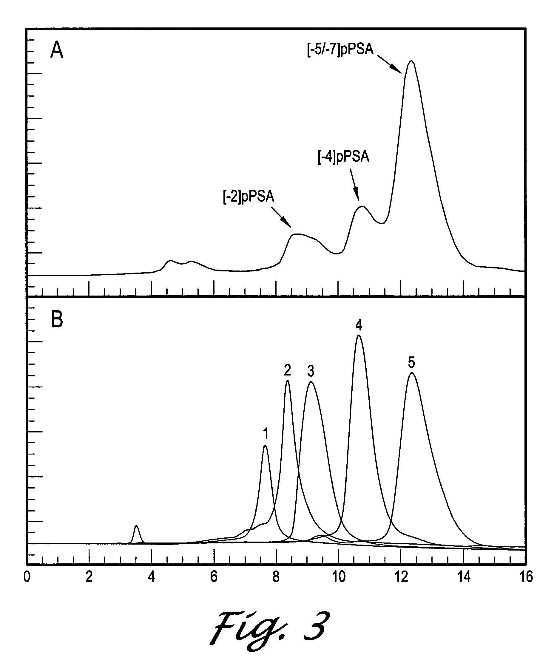
[0109]The presence of pPSA in human serum would indicate the following. First, that PSA is secreted as the pPSA form in human tissue and is converted to mature PSA extracellularly. Second, that pPSA is stable in human serum and thus may be a useful diagnostic marker for pCa or BPH. We evaluated the presence of pPSA in human serum by first using affinity purification to purify all forms of PSA present in a pool of human serum. We next fractionated the eluted PSA forms on HPLC and identified each PSA form based on its elution profile from the column. This analysis indicated that pPSA is present in human serum.

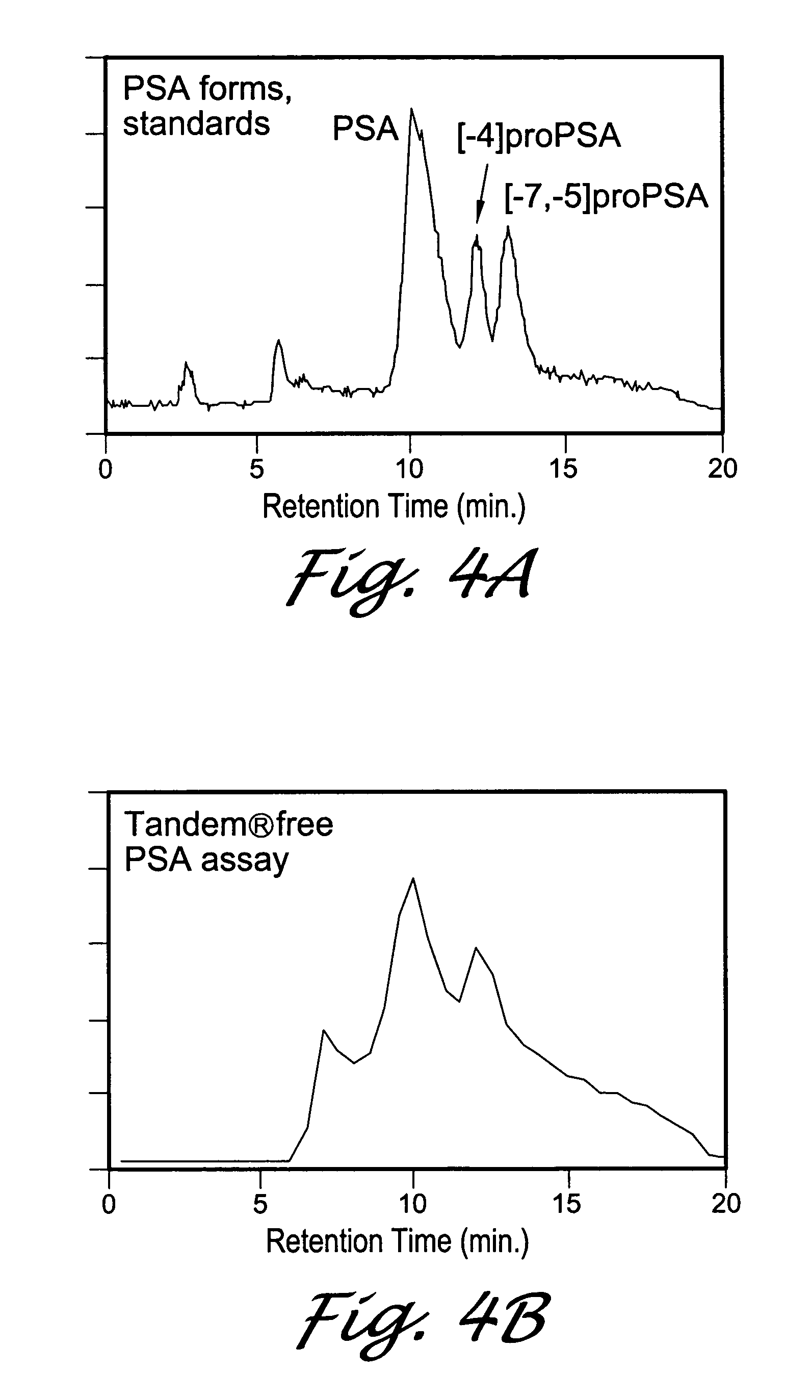
[0110]FIG. 4A shows the RT of standards of the different forms of PSA. All forms were verified by amino acid sequencing. The [−7, −5]pPSA peak contains approximately equal levels of [−7]pPSA and [−5]pPSA forms which are not resolved from each other. FIG. 4B shows the profile of PSA forms from serum bound to the PSM773 affinit...

example 3

Detection of Truncated pPSA Forms in Prostate Cancer Serum

[0112]Serum from several individual men were also analyzed by Western blot using newly devleoped pPSA mAbs. PSA was purified from the serum of men who were biopsy-positive and biopsy-negative for prostate cancer. The serum PSA values of the 5 biopsy-positive men were 6, 9, 10, 18, and 24 ng / ml. Three biopsy-negative men with 7, 10, and 12 ng / ml total PSA were also analyzed. Since the free PSA represented only 10-20% of the total PSA, and it was not known what percentage of the free PSA might be comprised of pPSA forms, it was necessary to purify PSA from 100-200 mls of serum in order to be assured of adequate detection sensitivity for Western blot analysis. Total PSA was purified from the serum by immunoaffinity chromatography using the anti-PSA mAb, PSM773, which recognizes all forms of free PSA and PSA bound to ACT. The recovery of PSA ranged from 30-60% of the amounts calculated to be in the starting serum by immunoassay. ...


PUM

PropertyMeasurementUnit
pHaaaaaaaaaa
pHaaaaaaaaaa
lengthaaaaaaaaaa

Abstract

Inactive precursor forms of PSA (pPSA) have been identified to exist in serum and tissues of patients with prostate cancer. Antibodies specific for pPSA are provided. Methods for detecting inactive precursors of PSA in human physiological fluid and tissues are also provided, as well as diagnostic kits and methods useful in the diagnosis and management of prostate cancer.

Description

[0001]This is a continuation-in-part of application Ser. No. 09 / 302,965, filed on Apr. 30, 1999, now abandoned, which in turn is a continuation-in-part of application Ser. No. 09 / 251,686, filed on Feb. 17, 1999, now abandoned, which in turn is a continuation of application Ser. No. 08 / 846,408, filed on Apr. 30, 1997, now abandoned. The content of the applications Ser. Nos. 09 / 251,686, 09 / 302,965, and 08 / 846,408 are incorporated herein in their entirety by reference.FIELD OF THE INVENTION[0002]The present invention relates generally to the detection and identification of proteins, as well as various forms and subunits of proteins, which have the potential utility as diagnostic markers. In particular, the present invention relates to the detection of inactive precursor forms of prostate specific antigens, and antibodies that are capable of preferentially binding to precursor forms of prostate specific antigens.BACKGROUND OF THE INVENTION[0003]The measurement of serum prostate specific...

Claims


Application Information

Patent Timeline
Patent Type & Authority Patents(United States)
IPC IPC(8): G01N33/53G01N33/574
CPCG01N33/57434
Inventor MIKOLAJCZYK, STEPHEN D.RITTENHOUSE, HARRY G.WANG, TANG JANGWOLFERT, ROBERT L.
Owner HYBRITECH
Why Eureka
Try Eureka