Computer aided disease detection system for multiple organ systems

a disease detection and computer-aided technology, applied in the field of digital medical image processing, can solve the problems of increasing the demands and difficulty of physician interpretation of images, increasing the shear quantity of image data, and affecting the accuracy of diagnosis, so as to reduce the reading time

Active Publication Date: 2010-03-02
PHILIPS HEALTHCARE INFORMATICS INC
View PDF11 Cites 30 Cited by
  • Summary
  • Abstract
  • Description
  • Claims
  • Application Information

AI Technical Summary

Benefits of technology

[0014]The present invention discloses a computer-aided disease detection system for multiple organ systems that can reduce the reading time for large-scale anatomical imaging studies of, e.g., abdomen or thorax, both two-and three-dimensional.

Problems solved by technology

However, such advances incur human and financial costs, particularly in terms of the cost of imaging studies and the necessary professional medical expertise needed to utilize and interpret the resultant imagery.
In the past decade, the rapid increase in three-and four-dimensional modalities (for example, computed tomography (CT), magnetic resonance imaging (MRI), single photon emission tomography (SPECT), and various others, along with rapid time sequences of such imaging operations) has led to an increase in the shear quantity of image data, and a concomitant increase in the demands and difficulty of physician interpretation of the imagery leading to subsequent diagnosis.
Some believe that the practice of clinical radiology is approaching a “crisis” time in which the proliferating quantity and categories of medical imaging data are threatening to overwhelm the available professional interpretive skills necessary for its exploitation.
One impediment to the widespread acceptance of CAD systems concerns the legal and ethical responsibility enjoined upon the medical professional to discern incidental findings—that is, to detect abnormal pathologies that are outside the primary focus of the study.
Thus, the ability for such CAD systems to substantially reduce interpretation time is greatly restricted.

Method used


Image

  • Computer aided disease detection system for multiple organ systems
  • Computer aided disease detection system for multiple organ systems
  • Computer aided disease detection system for multiple organ systems

Examples


Embodiment Construction

[0025]The following is a detailed description of the preferred embodiments of the invention, reference being made to the drawings in which the same reference numerals identify the same elements of structure in each of the several figures.

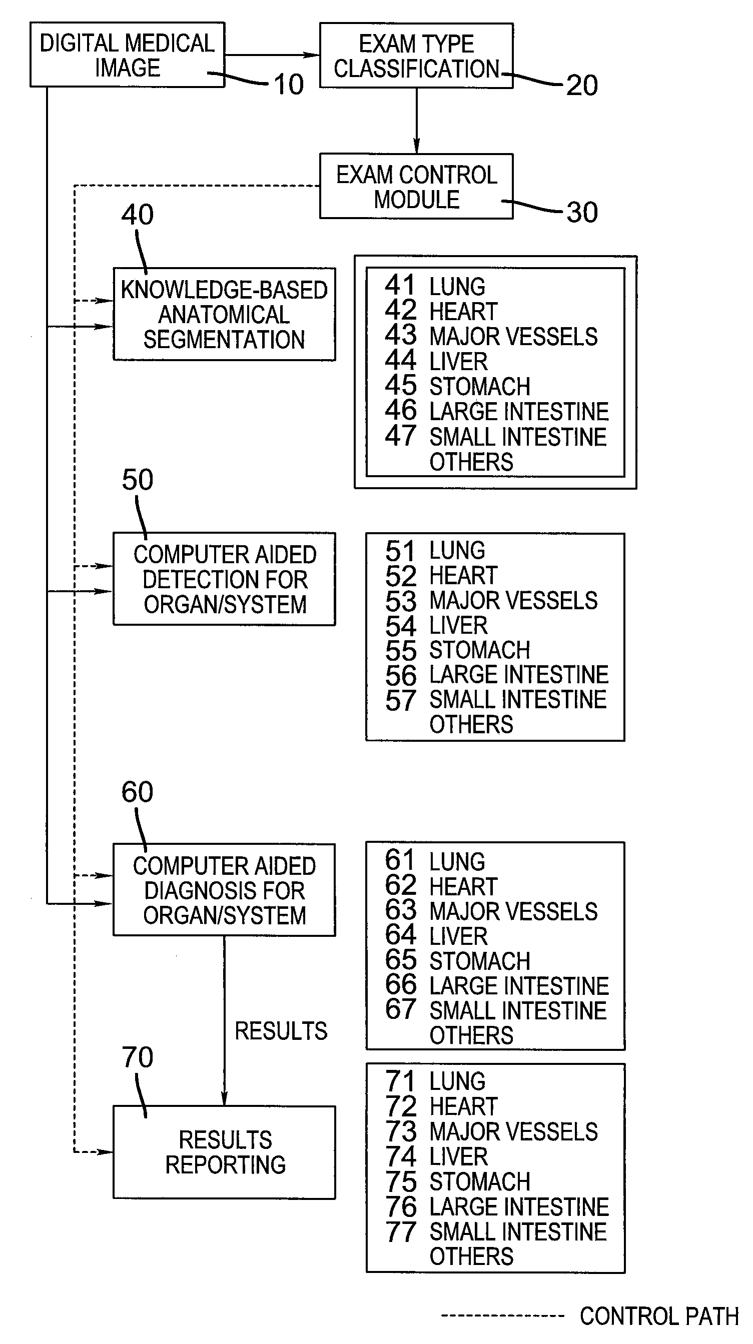
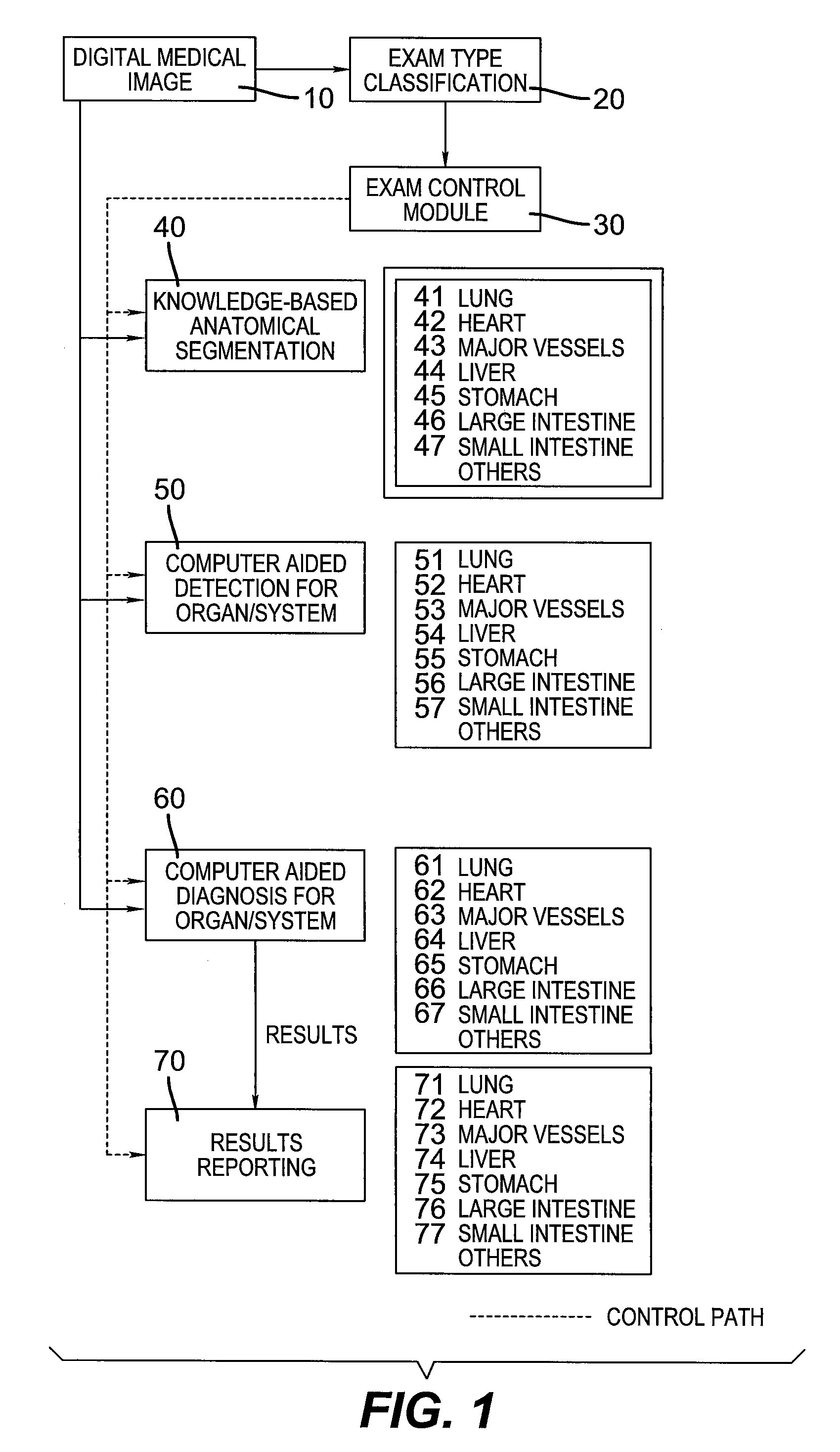
[0026]FIG. 1 shows a block diagram of the present invention. With reference to this figure, a digital medical image 10 is presented to the method. This image can be created by any of the well-known methods currently practiced in the medical field, e.g. computerized tomography. The image is preferably represented in the industry standard DICOM format.

[0027]A first stage of processing involves an optional exam-type classification step 20. The purpose of this step is to determine the specific patient anatomy that is present in the digitized image so as to direct the remaining processing to the specific organs or organ systems that are present in the image. For example, thoracic CT images contain information about patient lungs, heart, and major blood v...


PUM


Abstract

A computer aided disease detection system and method for multiple organ systems. The method performs computer aided examination of digital medical images. A patient exam type of a digital medical image is determined. Based on the patient exam type, one or more of a plurality of knowledge based anatomical segmentation blocks are invoked, each block performing image segmentation for a single organ or organ system present in the image. Based on the patient exam type, for each successfully segmented organ or organ system, one or more of a plurality of knowledge based computer aided detection blocks are invoked, each block of which is designed to search for and locate potential disease foci in a particular organ or organ system.

Description

CROSS REFERENCE TO RELATED APPLICATIONS[0001]Reference is made to, and priority is claimed from, commonly assigned Provisional Application No. 60 / 754,883, entitled “COMPUTER AIDED DISEASE DETECTION SYSTEM FOR MULTIPLE ORGAN SYSTEMS”, filed on Dec. 29, 2005 in the name of Nicponski, and which is incorporated herein by reference.FIELD OF THE INVENTION[0002]The invention relates generally to the field of digital medical image processing, and in particular to a method and system for automated computer examination of medical imagery for the purpose of detecting likely disease sites in multiple human organ systems.BACKGROUND OF THE INVENTION[0003]Technological advances in the field of non-invasive imaging of the human body have produced gains in the ability of medical professionals to detect, diagnose, and treat diseases, thereby leading to improved outcomes and reduced morbidity and mortality. Advance in imaging resolution, functionality, and quality lead to new applications of the vario...

Claims


Application Information

Patent Timeline
Patent Type & Authority Patents(United States)
IPC IPC(8): G06K9/00A61B5/00
CPCG06T7/0012
Inventor NICPONSKI, HENRY
Owner PHILIPS HEALTHCARE INFORMATICS INC
Why Eureka
Try Eureka