Method and apparatus for detecting anatomic structures

a technology of anatomic structure and method, applied in the field of medical diagnostic systems, can solve the problems of user's inability to precisely identify the location desired, system's inability to allow users to input precise measurements, and limited time with which to conduct measurements

Active Publication Date: 2010-03-16
GENERAL ELECTRIC CO
View PDF5 Cites 53 Cited by
  • Summary
  • Abstract
  • Description
  • Claims
  • Application Information

AI Technical Summary

Problems solved by technology

In ultrasound, due to patient and system limitations, the user is often unable to precisely identify the location desired, such as the endocardium or epicardium.
For example, the systems do not have the capability to allow the user to input precise measurements to track the minute variable contours of the tissue.
In addition, the user has limited time with which to conduct the measurements.
This is time consuming and the model assumes that the ventricle is symmetric along the diameter.
Also, due to poor image quality in many patients, locating the volume automatically can be difficult.
Other imaging modalities also acquire cardiac images and experience the same problems.

Method used


Image

  • Method and apparatus for detecting anatomic structures
  • Method and apparatus for detecting anatomic structures
  • Method and apparatus for detecting anatomic structures

Examples


Embodiment Construction

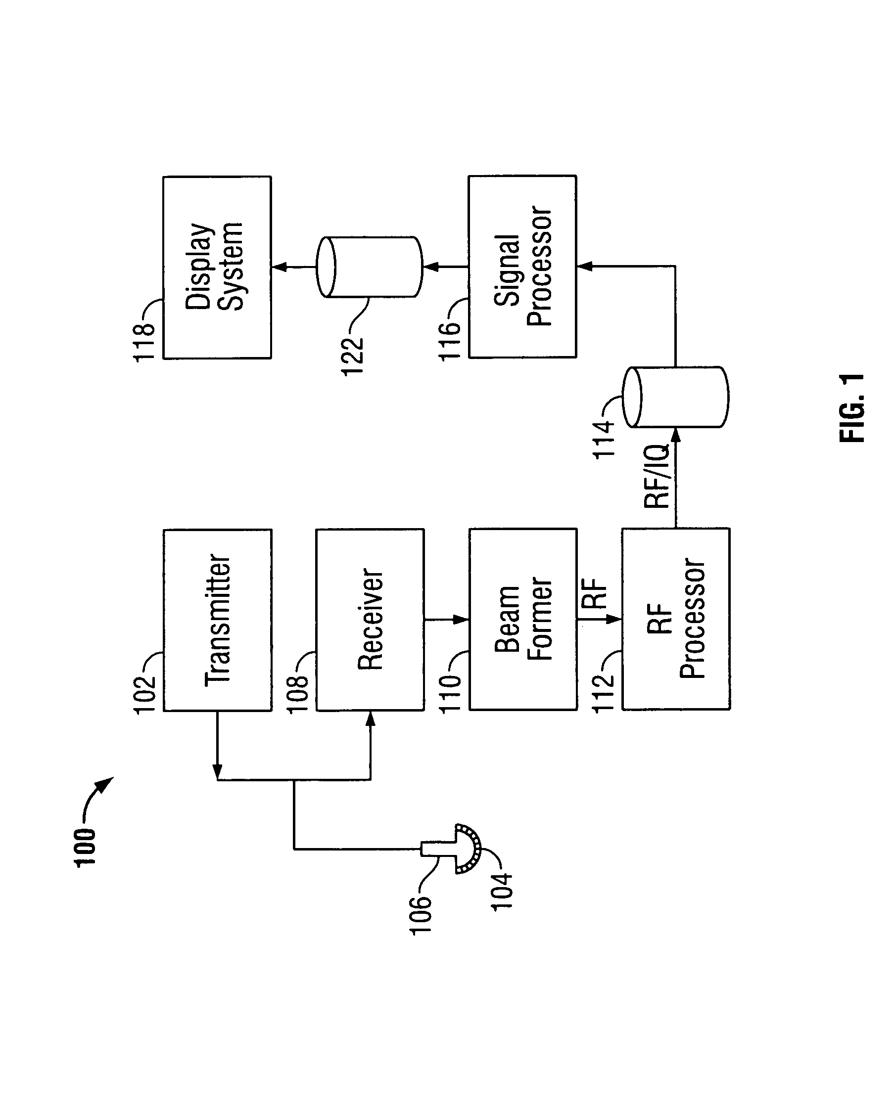
[0019]FIG. 1 illustrates a block diagram of an ultrasound system 100 formed in accordance with an embodiment of the present invention. The ultrasound system 100 includes a transmitter 102 which drives an array of elements 104 within a transducer 106 to emit pulsed ultrasonic signals into a body. A variety of geometries may be used. The ultrasonic signals are back-scattered from structures in the body, like blood cells or muscular tissue, to produce echoes which return to the elements 104. The echoes are received by a receiver 108. The received echoes are passed through a beamformer 110, which performs beamforming and outputs an RF signal. The RF signal then passes through an RF processor 112. Alternatively, the RF processor 112 may include a complex demodulator (not shown) that demodulates the RF signal to form IQ data pairs representative of the echo signals. The RF or IQ signal data may then be routed directly to RF / IQ buffer 114 for temporary storage.

[0020]The ultrasound system 1...


PUM


Abstract

An anatomic structure is detected based on a medical diagnostic imaging data set. The anatomic structure comprises at least two different types of tissue. At least one anatomic landmark is identified within the data set, the data set is overlaid with a contour template, and a search region of the data set surrounding the contour template is scanned to identify transition points associated with a predefined characteristic of the anatomic structure.

Description

BACKGROUND OF THE INVENTION[0001]This invention relates generally to medical diagnostic systems. In particular, the present invention relates to methods and apparatus for acquiring and processing diagnostic data sets to identify the location of the transition between different types of tissue and between tissue and blood.[0002]The processing of cardiac image data is of great interest to determine how the heart is functioning. Currently, many processing algorithms rely on the user to identify the data to be processed. In ultrasound, due to patient and system limitations, the user is often unable to precisely identify the location desired, such as the endocardium or epicardium. For example, the systems do not have the capability to allow the user to input precise measurements to track the minute variable contours of the tissue. In addition, the user has limited time with which to conduct the measurements.[0003]For example, an important measurement for left ventricular function is the ...

Claims


Application Information

Patent Timeline
Patent Type & Authority Patents(United States)
IPC IPC(8): A61B8/00A61B5/05G06F9/00G01T1/161A61B5/055A61B6/00A61B6/03A61B8/08A61B19/00G01T1/164G06T5/00
CPCA61B8/08G06T7/0083A61B6/503A61B8/0883G06T2207/10132G06T2207/30048G06T7/12
Inventor TORP, ANDERS HERMANRABBEN, STEIN INGEOLSTAD, BJORN
Owner GENERAL ELECTRIC CO
Why Eureka
Try Eureka