Method and apparatus for classifying tissue using image data

a tissue and image data technology, applied in the field of tissue classification, can solve the problems of only one parameter examination at a time, non-invasive magnetic resonance imaging (mri), and unsuitable detection of lymph node metastases

Active Publication Date: 2010-05-18
SIEMENS MEDICAL SOLUTIONS USA INC
View PDF6 Cites 12 Cited by
  • Summary
  • Abstract
  • Description
  • Claims
  • Application Information

AI Technical Summary

Problems solved by technology

Magnetic resonance imaging (MRI) is a non-invasive technique for providing images with excellent anatomical detail and soft tissue contrast, but is generally unsuitable for detection of lymph-node metastases.
However, the main drawback of this simple linear analysis is the fact that parameters can only be examined one at a time.

Method used


Image

  • Method and apparatus for classifying tissue using image data
  • Method and apparatus for classifying tissue using image data
  • Method and apparatus for classifying tissue using image data

Examples


Embodiment Construction

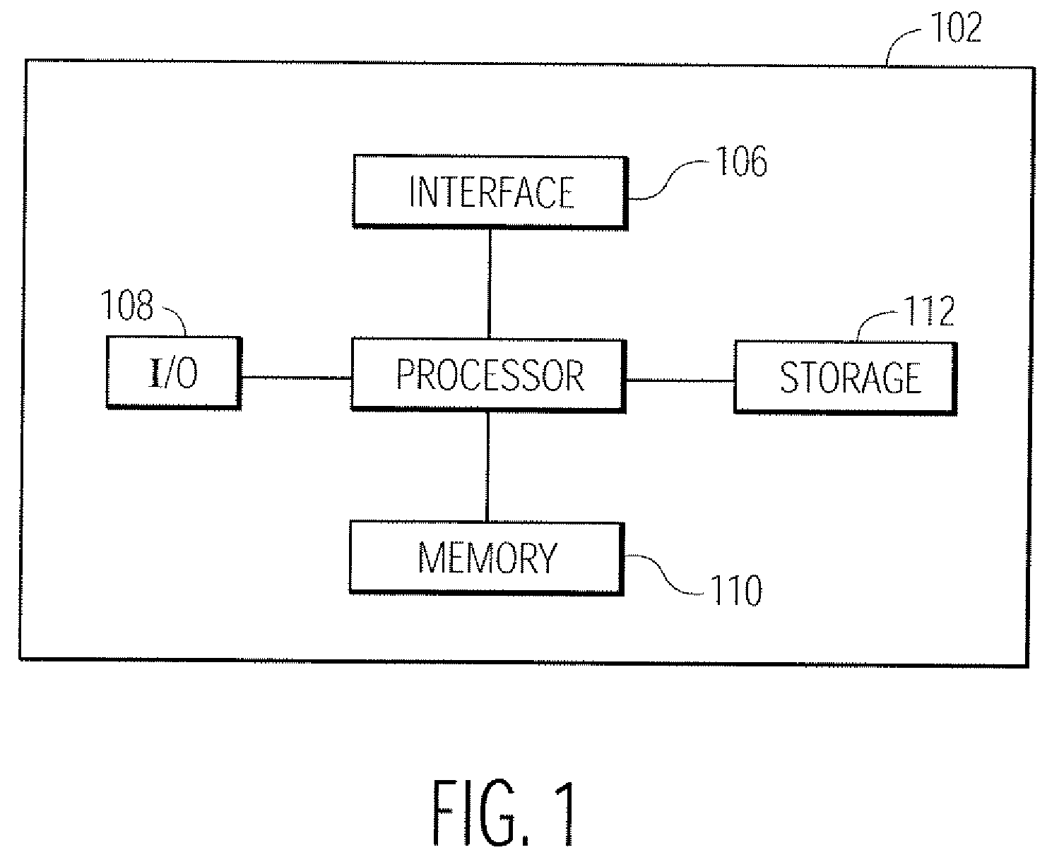
[0029]The processing steps described herein may be performed by an appropriately programmed computer, the configuration of which is well known in the art. An appropriate computer may be implemented, for example, using well known computer processors, memory units, storage devices, computer software, and other components. A high level block diagram of such a computer is shown in FIG. 1. Computer 102 contains a processor 104 which controls the overall operation of computer 102 by executing computer program instructions which define such operation. The computer program instructions may be stored in a storage device 112 (e.g., magnetic disk, optical disk, or any other computer readable medium) and loaded into memory 110 when execution of the computer program instructions is desired. Memory 110 may also be used to store data used during the various steps of the method. Computer 102 also includes one or more interfaces 106 for communicating with other devices (e.g., locally or via a networ...


PUM


Abstract

Disclosed is a technique for classifying tissue based on image data. A plurality of tissue parameters are extracted from image data (e.g., magnetic resonance image data) to be classified. The parameters are preprocessed, and the tissue is classified using a classification algorithm and the preprocessed parameters. In one embodiment, the parameters are preprocessed by discretization of the parameters. The classification algorithm may use a decision model for the classification of the tissue, and the decision model may be generated by performing a machine learning algorithm using preprocessed tissue parameters in a training set of data. In one embodiment, the machine learning algorithm generates a Bayesian network. The image data used may be magnetic resonance image data that was obtained before and after the intravenous administration of lymphotropic superparamagnetic nanoparticles.

Description

[0001]This application claims the benefit of U.S. Provisional Application No. 60 / 699,805 filed Jul. 15, 2005, which is incorporated herein by reference.BACKGROUND OF THE INVENTION[0002]The present invention relates generally to tissue classification, and more particularly to classifying a pathological state of tissue based on image data.[0003]Accurate detection of tissue metastases is essential in optimizing treatment for solid cancers. In particular, there is a need for non-invasive techniques for identifying such metastases. The following description will generally focus on detection of lymph-node metastases, but it should be understood that the present invention is not so limited, and can be applied to any type of tissue.[0004]Magnetic resonance imaging (MRI) is a non-invasive technique for providing images with excellent anatomical detail and soft tissue contrast, but is generally unsuitable for detection of lymph-node metastases. However, the quality of MRI can be improved by u...

Claims


Application Information

Patent Timeline
Patent Type & Authority Patents(United States)
IPC IPC(8): G06K9/00A61B5/05G06V10/84
CPCG06K9/00147G06T7/0012G06K9/6296G06T2207/30024G06V20/698G06V10/84G06F18/29
Inventor FUCHS, THOMASWACHMANN, BERNDNEUBAUER, CLAUSCHENG, JIE
Owner SIEMENS MEDICAL SOLUTIONS USA INC
Why Eureka
Try Eureka