Endoscopic attachment device

a technology of endoscope and attachment device, which is applied in the field of endoscope, can solve the problems of device not allowing visualisation during medical procedure, unsatisfactory surgical procedure, and great discomfort for patients, and achieves excellent template and matrix, greater resistance, and greater efficiency.

Active Publication Date: 2010-07-20
AXONICS WOMENS HEALTH LTD
View PDF20 Cites 51 Cited by
  • Summary
  • Abstract
  • Description
  • Claims
  • Application Information

AI Technical Summary

Benefits of technology

[0024]It is an object of the present invention to provide an endoscopic device that allows for the surgeon to manipulate the working element during operation and / or diagnosis while keep non-working elements immobile.
[0100]In a further embodiment of the invention, the device prosthetic device comprises cells, such as stem cells or fat cells. Polyacrylamide provides an excellent template and matrix for cell growth. The use of cells with the hydrogel of the invention for the preparation of the device would allow for cellular engraftment to the surrounding tissue in the ureter, urethra or analis canalis. A device comprising the hydrogel of the invention and the appropriate cells allows for greater resistance and greater efficiency in providing resistance.

Problems solved by technology

Devices cumbersome to handle or manipulate may result in unsuccessful surgical procedures, misdiagnoses and, particularly in the case where the patient is not anaesthetised, great discomfort to the patient.
Moreover, the device does not allow for visualisation during the medical procedure.
The device does not allow for variable positions of injection nor for visualisation during the medical procedure.

Method used


Image

  • Endoscopic attachment device
  • Endoscopic attachment device
  • Endoscopic attachment device

Examples


Embodiment Construction

[0024]It is an object of the present invention to provide an endoscopic device that allows for the surgeon to manipulate the working element during operation and / or diagnosis while keep non-working elements immobile.

[0025]Further, it is an object of the present invention to provide for a sheath of an endoscopic equipment which needs minimal sterilisation and is disposable.

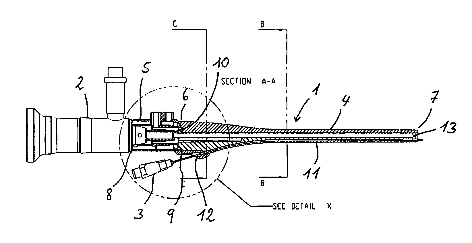
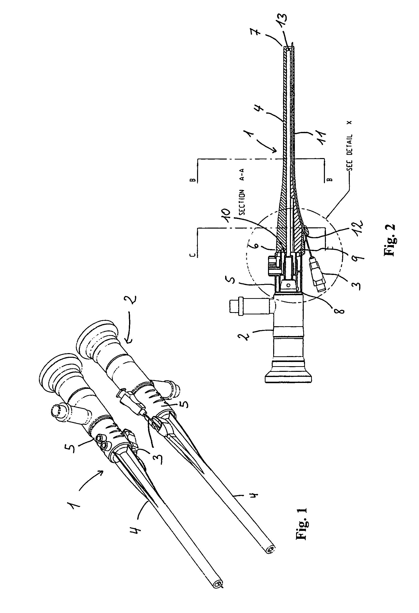
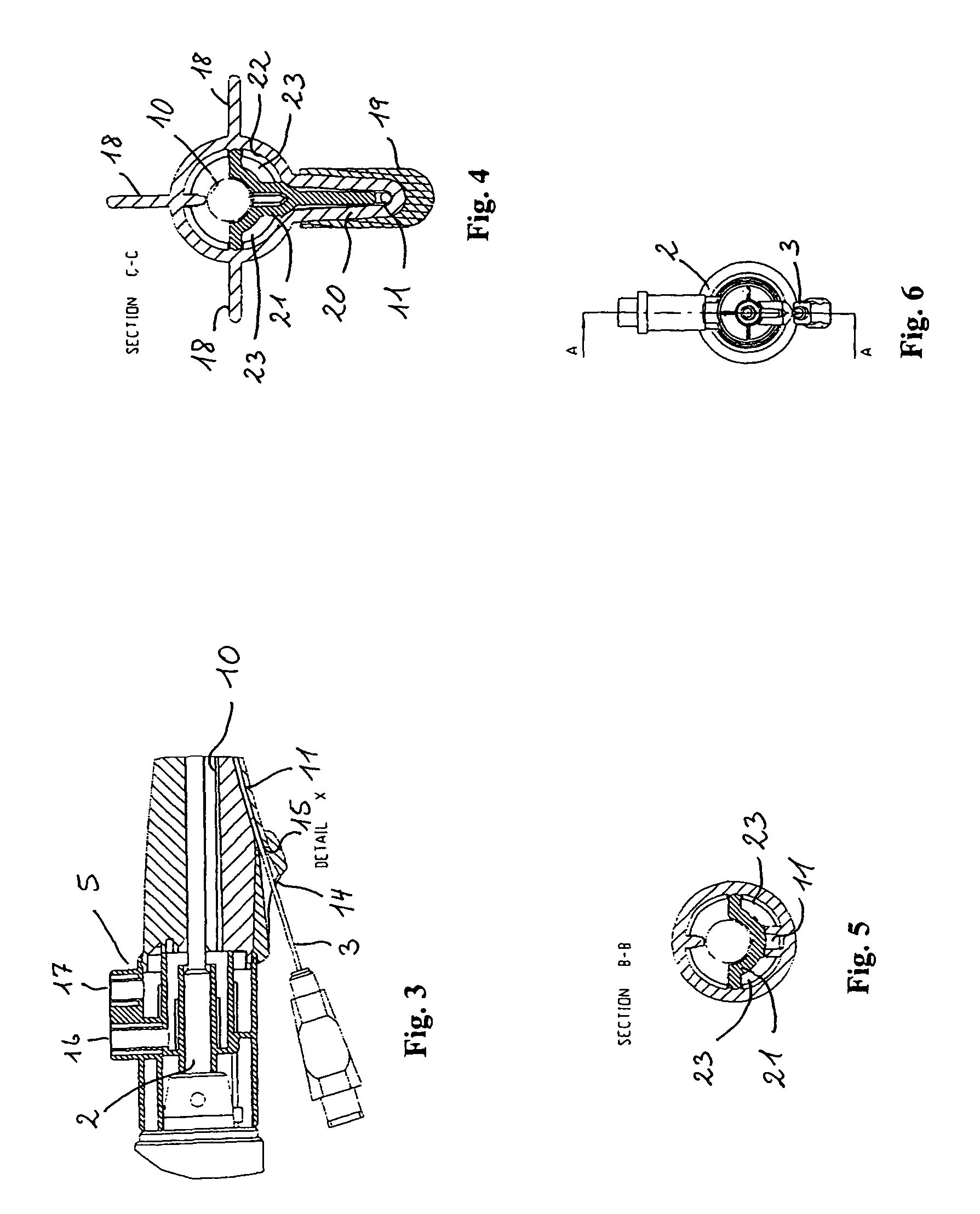
[0026]According to the invention, the above objectives is fulfilled by a sheath device suitable for endoscopic instruments, the device comprising:[0027]a) an elongated tubular member comprising:[0028]a proximal end,[0029]a distal open end, and[0030]at least one fluid channel extending longitudinally from said proximal end to a fluid exit; and[0031]b) a flushing unit connected to said proximal end of the tubular member and comprising:[0032]a proximal open end suitable for receiving a first endoscopic instrument,[0033]a fluid inlet being in contact with said fluid channel, and[0034]a fluid outlet,[0035]the tubular me...


PUM


Abstract

A sheath device (1) suitable for endoscopic instruments (2) and comprising an elongated tubular member (4) having a proximal end (6), a distal open end (7), and at least one fluid channel (23). A flushing unit (5) is connected to the proximal end (6) of the tubular member (4) and comprises a proximal open end (8) suitable for receiving a first endoscopic instrument (2), a fluid inlet (16) in contact with said fluid channel (23), and a fluid outlet (17). The tubular member and the flushing unit together define a first interior guiding passage (10) for sheathing at least a part of said first endoscopic instrument (2). The connection between the tubular member (4) and the flushing unit (5) allows for axial rotation of the tubular member (4) in relation to the flushing unit (5).

Description

CROSS-REFERENCE TO RELATED APPLICATIONS[0001]The present application claims priority to U.S. Provisional Patent Application No. 60 / 479,725 filed Jun. 20, 2003, which is hereby incorporated by reference in its entirety.FIELD OF THE INVENTION[0002]The present invention relates to a sheath device suitable for an endoscopic device, such as a catheter or cystoscope, which, said sheath when mounted onto any optical device, allows for the surgeon to examine or manipulate within an orifice or through a incision, whilst easily manipulating the endoscopic device. The device allows for delivery by means of an agent to the target organ or tissue at variable positions whilst maintaining a fixed position of the optical device or fluid inlet / outlet device.BACKGROUND OF THE INVENTION[0003]In medical practise, whether for purposes of examination or surgical procedures, the devices used in the practice must allow for the surgeon to easily interact with the patient's organs and cavities. Devices cumbe...

Claims


Application Information

Patent Timeline
Patent Type & Authority Patents(United States)
IPC IPC(8): A61B1/00A61B1/12
CPCA61B1/00094A61B1/00105A61B1/00135A61B1/00142A61B1/12A61B17/3478A61B2017/00805A61B2017/345
Inventor HEM, SOREN
Owner AXONICS WOMENS HEALTH LTD
Why Eureka
Try Eureka