
Endoscopic resection method

a mucosal resection and endoscopic technology, applied in the field of methods for removing tissue from patients, can solve the problems of increasing patient discomfort, unnecessary burning or trauma beyond the desired treatment site, and into the surrounding anatomical lumen wall,

Active Publication Date: 2010-09-07
BOSTON SCI SCIMED INC
View PDF13 Cites 13 Cited by
  • Summary
  • Abstract
  • Description
  • Claims
  • Application Information

AI Technical Summary

Benefits of technology

The present invention provides methods for removing tissue from a patient's body that overcome limitations and disadvantages of previous methods. The methods involve injecting fluid into the submucosal layer to raise the targeted tissue and positioning electrodes to apply current through the tissue. The methods may include using a conductive snare loop or a combination of snare loops, and may also involve using a flexible sheath and injecting the fluid through an injection needle. The methods may be used in an endoscopic procedure or in a placement catheter. The technical effects of the invention include improved precision and control over tissue removal, reduced bleeding and risk of complications, and improved access to difficult-to-reach tissue.

Problems solved by technology

While useful in stemming the flow of blood resulting from tissue resection, electrosurgical methods can often cause unnecessary burning or trauma beyond the desired treatment site and into the surrounding anatomical lumen wall during a resection procedure.
As physicians sometimes use different devices to perform different functions, for example, use one catheter to make an incision and another to perform hemostasis or irrigation, the exchange of catheters to provide different functions extends the time to complete therapy, increases the risk to the patient, and also increases patient discomfort.
Consequently, physicians have to weigh the time, complexity, and benefits of interchanging single or dual purpose catheters to change treatment modalities against whatever disadvantage may result by working with a single catheter.

Method used


Image

  • Endoscopic resection method
  • Endoscopic resection method
  • Endoscopic resection method

Examples


Embodiment Construction

[0025]Reference will now be made in detail to the present exemplary embodiments of the invention illustrated in the accompanying drawings. Wherever possible, the same reference numbers will be used throughout the drawings to refer to the same or like parts.

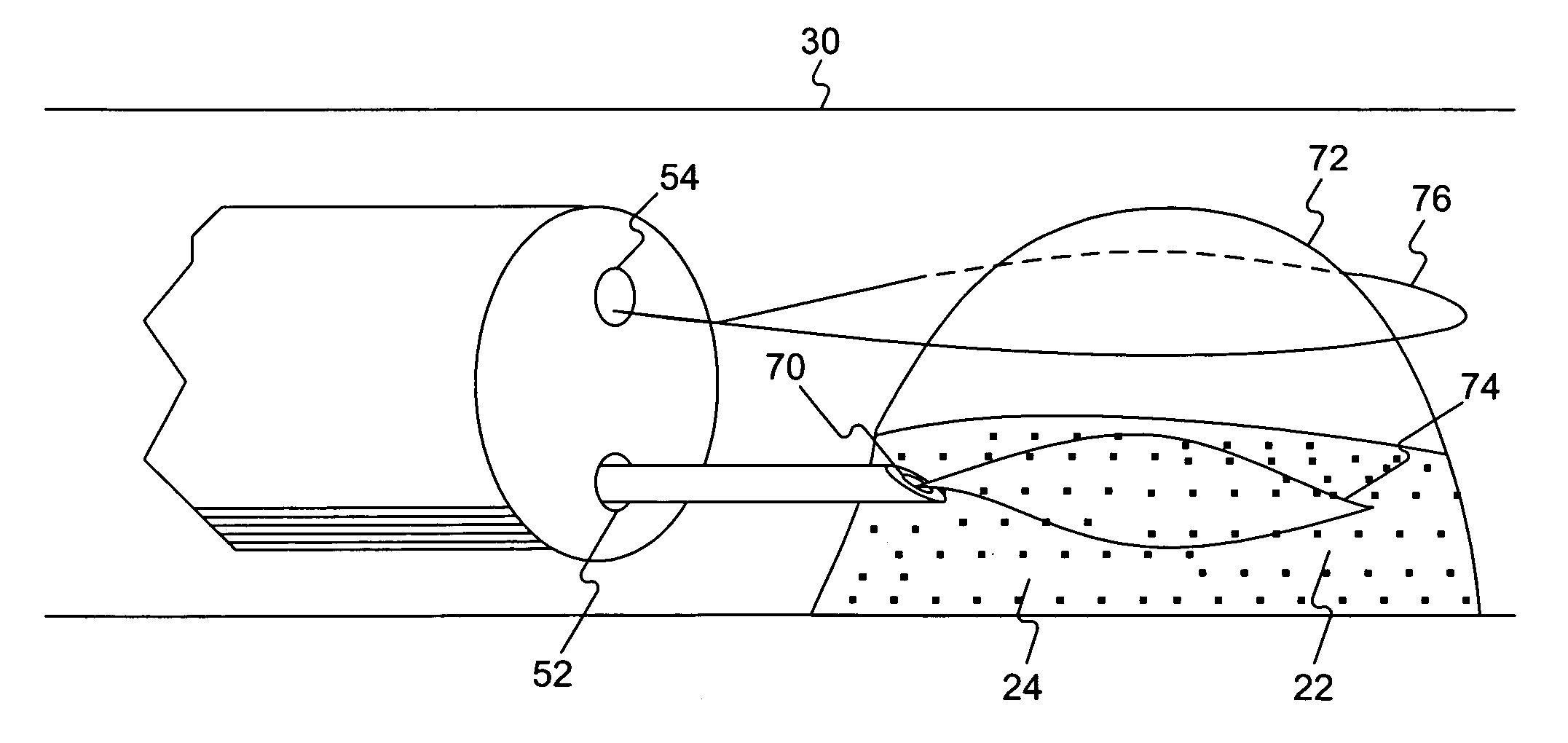
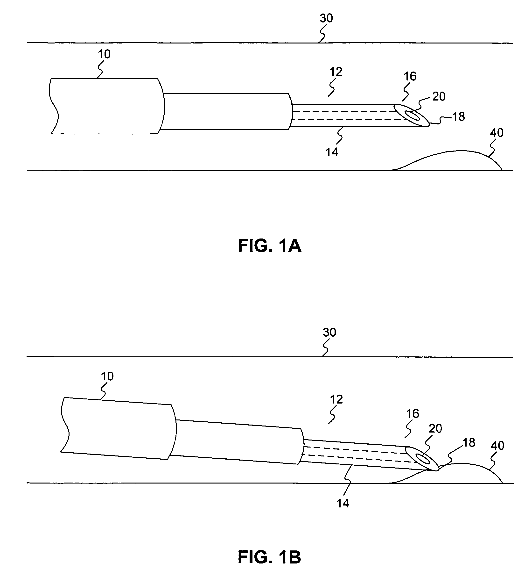
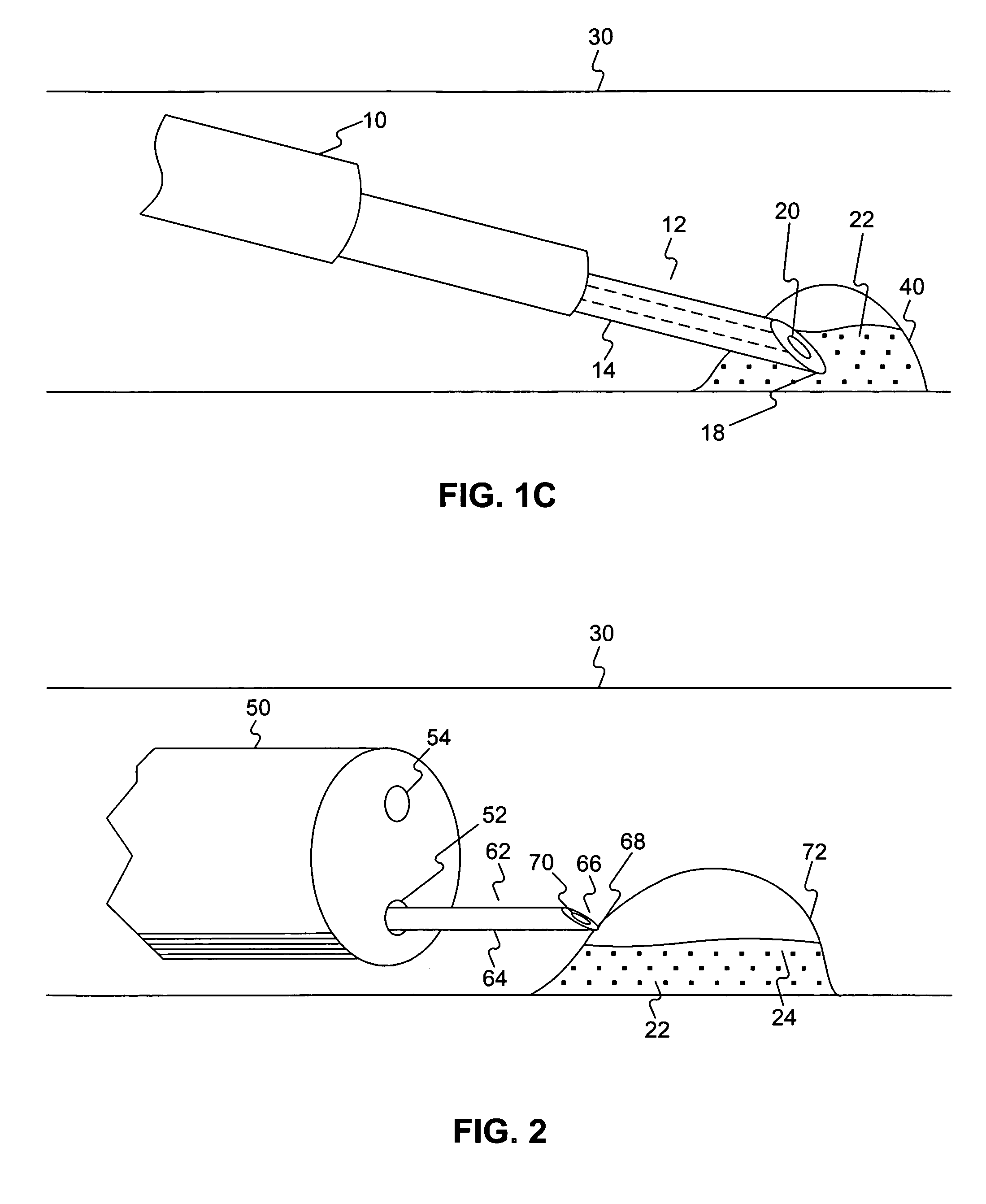
[0026]FIG. 1A illustrates an endoscope 10 positioned within a patient's anatomical lumen 30 for the treatment of a sessile adenoma 40. An injection needle 12 is positioned within the anatomical lumen 30 through a working channel of the endoscope 10. The injection needle 12 includes a shaft 14, a piercing tip 16 having a bevel 18 and an opening 20 at its distal end (i.e. the end further from the operator during use). During endoscopic treatment procedures, injection needles are often used, for example, to introduce irrigation fluids at a treatment site, inject vaso-constrictor fluid into a vessel to slow hemorrhaging, or inject a sclerosing agent to control bleeding varices by hardening the targeted tissue.

[0027]Referring to FIG. 1...


PUM


Abstract

Embodiments of the invention are directed to a method of excising tissue including injecting fluid into the submucosa to raise targeted tissue. A first electrode is positioned below the targeted tissue within the injected fluid and a second electrode is positioned adjacent a surface of the raised targeted tissue opposite the first electrode. Electrical current is applied between the first and second electrodes and the targeted tissue is excised with the second electrode.

Description

FIELD OF THE INVENTION[0001]Embodiments of this invention relate to methods for removing tissue from a patient. More particularly, at least certain embodiments of the invention relate to methods for endoscopic mucosal resection including injecting fluid into tissue, positioning wire snares at a treatment site, and applying current between the snares in order to cut and remove tissue.BACKGROUND OF THE INVENTION[0002]Endoscopic methods are commonly used for diagnosis and / or treatment of internal anatomical lumens within a patient's body, such as, for example, the gastrointestinal tract. For example, there are several methods, known as endoscopic mucosal resection, for treating both malignant and non-malignant tumors within a patient's mucosa (the mucous tissue lining various internal anatomical lumens consisting of epithelium, lamina, propria, and, in the gastrointestinal tract, a layer of smooth muscle). Endoscopic mucosal resection may include snaring and then excising sessile adeno...

Claims


Application Information

Patent Timeline
Patent Type & Authority Patents(United States)
IPC IPC(8): A61B18/12
CPCA61B18/14A61B2018/00482A61B2018/00601A61B2018/00982A61B2018/1407A61B2018/141A61B2218/002A61B2017/00269A61B18/1492A61B18/1482A61B18/16A61B2018/00589A61B2018/00595A61B2018/1467
Inventor WEITZNER, BARRY
Owner BOSTON SCI SCIMED INC
Learn More