Endoscope device

An endoscope and image signal technology, applied in the fields of endoscopy, medical science, surgery, etc., can solve the problems of wrong cutting, unclear boundary between muscularis propria and submucosa, and reduced visual recognition of blood vessels

Active Publication Date: 2013-04-24
OLYMPUS CORP
View PDF10 Cites 6 Cited by
  • Summary
  • Abstract
  • Description
  • Claims
  • Application Information

AI Technical Summary

Problems solved by technology

[0008] However, when the staining of the submucosa is too dark, the visibility of blood vessels decreases, and blood vessels in the deep mucosa may be mistakenly cut. Conversely, when the staining of the submucosa is too light, the visibility of blood vessels increases, but the muscularis propria is present. Problems with blurring of the border with the submucosa

Method used


Image

  • Endoscope device
  • Endoscope device
  • Endoscope device

Examples


Deformed example 1

[0097] This modified example is a modified example of the light source device. Figure 17 It is a configuration diagram showing the configuration of an endoscope apparatus according to the modification. Figure 17 and figure 1 The structure of the light source device is different. exist Figure 17 in, for figure 1 The same structural elements are marked with the same reference numerals and descriptions are omitted.

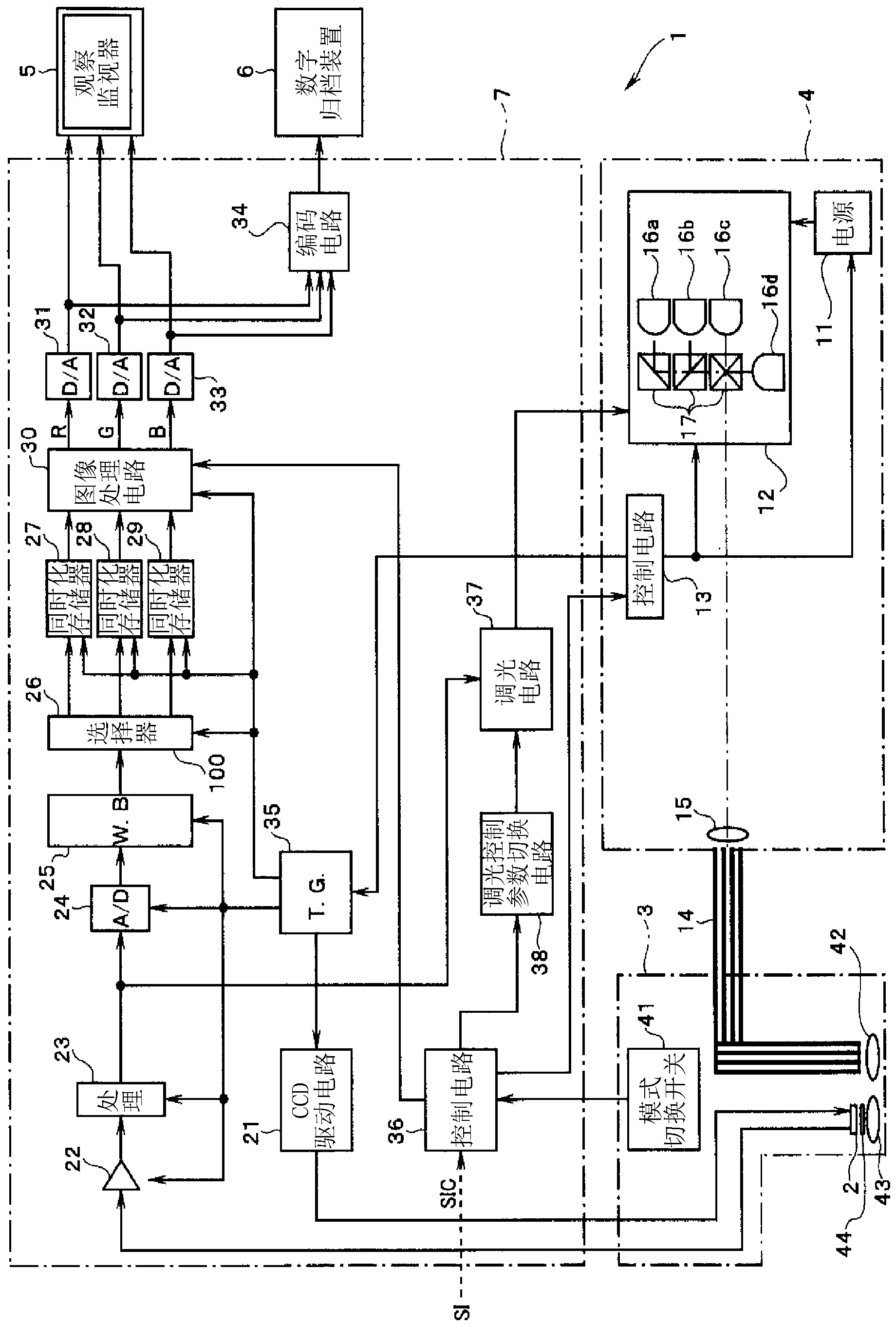
[0098] The light source device 4A is configured to include: a xenon lamp 101 that emits illumination light (white light) as an illumination unit; an infrared cut filter 102 that cuts off the white light; and a diaphragm that controls the amount of white light passing through the infrared cut filter 102 device 103; rotary filter 104 as a wavelength band limiting unit that makes illumination light into surface sequential light; and control circuit 13A that controls the rotation and position of rotary filter 104. The surface sequential light passing through the ...

Deformed example 2)

[0112] In the above-mentioned embodiments and modifications, the narrowband light NBa with a wavelength of around 630 nm was used to draw relatively thick blood vessels. However, narrowband light NBc with a wavelength of around 550 nm may be used to draw thinner blood vessels. That is, the narrowband light NBc around a wavelength of 550 nm is light in a wavelength band on the shorter wavelength side than the narrowband light NBb around a wavelength of 600 nm, and is less likely to be affected by scattering or absorption by the tissue surface in the body cavity.

[0113] The narrow-band light NBc with a wavelength around 550nm has the advantages of being less susceptible to the influence of indigo carmine and capable of depicting thinner blood vessels.

[0114] As described above, also with the structure shown in this modified example, the same actions and effects as those of the above-mentioned embodiment can be obtained.

Deformed example 3)

[0116] In the above-described embodiment, the composite image CP including the fluorescence image FP is displayed in the ESD mode, but the fluorescence image FP may not be displayed in the composite image CP in the ESD mode.

[0117] In this modified example, the ESD mode further includes a fluorescence image display mode FM in which the fluorescence image FP is displayed in the composite image CP, and a fluorescence image non-display mode NFM in which the fluorescence image FP is not displayed in the composite image CP.

[0118] In ESD, when a spark or the like is generated by use of an electric knife or the like, smudges or halos may be generated on an observation image displayed on the observation monitor 5 . Since autofluorescence FL is originally weak light, when the magnification of autofluorescence FL is high and halos or the like are generated, the observation image displayed on the observation monitor 5 may be greatly disturbed, and observation may not be possible.

...


PUM


Abstract

An endoscope device (1), which comprises a light source (4), an endoscope (3), a processor (7) and a viewing monitor (5). The light source (4) switches the illumination light from an excitation light (EX) for exciting a connective tissue in the submucosa of a tissue within a body cavity to a narrowband light (NBa) that is absorbed by a blood vessel (64) running through the submucosa (62) or muscularis propria (63) and a narrowband light (NBb) that is absorbed by a substance topically injected into the submucosa (62). The processor (7) generates multiple image signals, said image signals having been obtained by taking images with CCD2 of the endoscope (3), and the viewing monitor (5) synthesizes and displays multiple images.

Description

technical field [0001] The present invention relates to endoscopic devices, and more particularly to endoscopic devices suitable for submucosal dissection. Background technique [0002] Conventionally, in the medical field, various low-invasive examinations and operations using endoscopes have been performed. The operator inserts the endoscope into the body cavity, observes the subject captured by the imaging device installed at the front end of the endoscope insertion part, and can treat the lesion with the treatment instrument inserted into the treatment instrument channel as needed. department for processing. Surgery using an endoscope does not require laparotomy or the like, and thus has the advantage of reducing the burden on the patient's body. [0003] The endoscope device is configured to include an endoscope, an image processing device connected to the endoscope, and an observation monitor. The lesion is imaged by an imaging element provided at the distal end of ...

Claims


Application Information

Patent Timeline
Patent Type & Authority Applications(China)
IPC IPC(8): A61B1/00
CPCA61B1/00009A61B1/0638A61B1/043A61B1/0669A61B1/0646A61B1/000094A61B1/0655
Inventor 武井俊二五十岚诚
Owner OLYMPUS CORP
Why Eureka
Try Eureka