
Engineered three-dimensional connective tissue constructs and methods of making the same

a three-dimensional connective tissue and construct technology, applied in the health care industry, can solve the problems of low rate of new therapeutic discovery, long drug discovery process, and high cost of new drug discovery, and achieve the effect of improving the efficiency of the process, improving the safety of patients, and improving the quality of li

Inactive Publication Date: 2014-04-10
ORGANOVO
View PDF4 Cites 57 Cited by
  • Summary
  • Abstract
  • Description
  • Claims
  • Application Information

AI Technical Summary

Benefits of technology

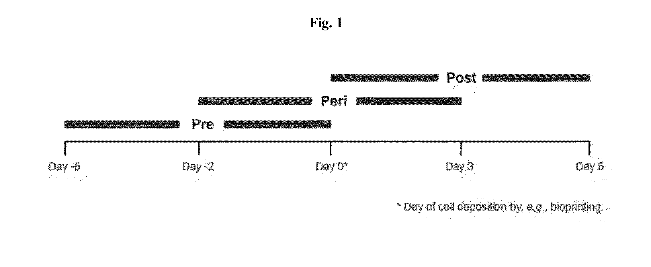
This patent describes a process for creating living, three-dimensional connective tissue constructs that are free of pre-formed scaffolds and are suitable for implantation in a subject. The process involves depositing multi-potent cells onto a support and exposing them to differentiation signals to create the desired tissue type. The constructs can also contain other cell types, such as vascular cells, to improve their suitability for implantation. The process can be performed using bioprinting technology. The resulting constructs can be used for various applications such as drug discovery, drug testing, toxicology testing, disease modeling, and cell screening.

Problems solved by technology

A number of pressing problems confront the healthcare industry.
Additionally, the research and development cost of a new pharmaceutical compound is approximately $1.8 billion.
Despite advances in technology and understanding of biological systems, drug discovery is still a lengthy, expensive, and inefficient process with low rate of new therapeutic discovery.

Method used


Image

  • Engineered three-dimensional connective tissue constructs and methods of making the same
  • Engineered three-dimensional connective tissue constructs and methods of making the same
  • Engineered three-dimensional connective tissue constructs and methods of making the same

Examples


example 1

MSC Culture

[0165]MSCs were cultured and expanded in standard cell culture conditions using a basal media that contained 5-10% (v:v) fetal bovine serum in low glucose DMEM supplemented with L-glutamine. In some cases, the MSCs were cultured in low (3-5%) oxygen conditions.

example 2

NovoGel™ Solutions and Mold

Preparation of 2% and 4% (w / v) NovoGel™ Solution

[0166]1 g or 2 g (for 2% or 4% respectively) of NovoGel™ (Organovo, San Diego, Calif.) was dissolved in 50 ml of Dulbecco's phosphate buffered saline (DPBS; Invitrogen Corp., Carlsbad, Calif.). Briefly, the DPBS and NovoGel™ are heated to 85° C. on a hot plate with constant stirring until the NovoGel™ dissolves completely. NovoGel™ solution is sterilized by steam sterilization at 125° C. for 25 minutes. The NovoGel™ solution remains in liquid phase as long as the temperature is maintained above 36.5° C. Below this temperature a phase transition occurs, the viscosity of the NovoGel™ solution increases and the NovoGel™ forms a solid gel.

Preparation of NovoGel™ Mold

[0167]A NovoGel™ mold was fabricated for the incubation of bio-ink (in the form of cellular cylinders) using a Teflon® mold that fit a 10 cm Petri dish. Briefly, the Teflon® mold was pre-sterilized using 70% ethanol solution and subjecting the mold to...

example 3

Fabrication of MSC-HAEC Bio-Ink

[0168]To prepare bio-ink (in the form of mixed cellular cylinders) MSC and HAEC were individually collected and then mixed at pre-determined ratios. Briefly, the culture medium was removed from confluent culture flasks and the cells were washed with DPBS (1 ml / 5 cm2 of growth area). Cells were detached from the surface of the culture flasks by incubation in the presence of trypsin (1 ml / 15 cm2 of growth area; Invitrogen Corp., Carlsbad, Calif.) for 10 minutes. MSC were detached using 0.15% trypsin while HAEC were detached using 0.1% trypsin. Following the incubation appropriate culture medium was added to the flasks (2× volume with respect to trypsin volume). The cell suspension was centrifuged at 200 g for 6 minutes followed by complete removal of supernatant solution. Cell pellets were resuspended in respective culture medium and counted using a hemocytometer. Appropriate volumes of MSC and HAEC were combined to yield a mixed cell suspension containi...


PUM

PropertyMeasurementUnit
time intervalsaaaaaaaaaa
heightaaaaaaaaaa
heightaaaaaaaaaa

Abstract

Disclosed are engineered, living, three-dimensional connective tissue constructs comprising connective tissue cells. In some embodiments, the connective tissue cells are derived from multi-potent cells such as mesenchymal stem / stromal cells. In some embodiments, the cells are cohered to one another. In some embodiments, the multi-potent cells have been exposed to one or more differentiation signals to provide a living, three-dimensional connective tissue construct. In some embodiments, the constructs are substantially free of pre-formed scaffold at the time of use. Also disclosed are implants for engraftment, arrays of connective tissue constructs for in vitro experimentation, as well as methods of making the same.

Description

CROSS-REFERENCE TO RELATED APPLICATIONS[0001]This application claims the benefit of U.S. application Ser. No. 61 / 661,768, filed Jun. 19, 2012, which is hereby incorporated by reference in its entirety.BACKGROUND OF THE INVENTION[0002]A number of pressing problems confront the healthcare industry. As of June 2012 there were 114,636 patients registered by United Network for Organ Sharing (UNOS) as needing an organ transplant. According to UNOS, between January and March 2012 only 6,838 transplants were performed. Each year more patients are added to the UNOS list than transplants are performed, resulting in a net increase in the number of patients waiting for a transplant.[0003]Additionally, the research and development cost of a new pharmaceutical compound is approximately $1.8 billion. See Paul, et al. (2010). How to improve R&D productivity: the pharmaceutical industry's grand challenge. Nature Reviews Drug Discovery 9(3):203-214. Drug discovery is the process by which drugs are di...

Claims


Application Information

Patent Timeline
Patent Type & Authority Applications(United States)
IPC IPC(8): C12N5/077
CPCC12N5/0654A61L27/3834A61L27/3886C12N5/0062C12N2502/1358C12N2502/28A61F2/08A61F2/28A61L27/56C12N5/0652G01N33/5008
Inventor PRESNELL, SHARON C.SHEPHERD, BENJAMIN R.EVINGER, III, ALBERT J.
Owner ORGANOVO
Learn More