
Cardiovascular risk assessments using aortic calcification information derived from x-ray measurements taken with a dual energy x-ray densitometer

aortic calcification technology, which is applied in the field of cardiac risk assessment using aortic calcification information derived from x-ray measurements taken with a dual-energy x-ray densitometer, to achieve the effects of reducing x-ray dose, shortening time, and increasing dos

Active Publication Date: 2010-09-28
HOLOGIC INC
View PDF7 Cites 5 Cited by
  • Summary
  • Abstract
  • Description
  • Claims
  • Application Information

AI Technical Summary

Problems solved by technology

While the images produced using x-ray bone densitometry IVA are not of sufficient quality for general radiological use, it has been observed that the quality of IVA images is similar to that of abdominal radiographs for visualizing abdominal aorta calcifications.

Method used


Image

  • Cardiovascular risk assessments using aortic calcification information derived from x-ray measurements taken with a dual energy x-ray densitometer
  • Cardiovascular risk assessments using aortic calcification information derived from x-ray measurements taken with a dual energy x-ray densitometer
  • Cardiovascular risk assessments using aortic calcification information derived from x-ray measurements taken with a dual energy x-ray densitometer

Examples


Embodiment Construction

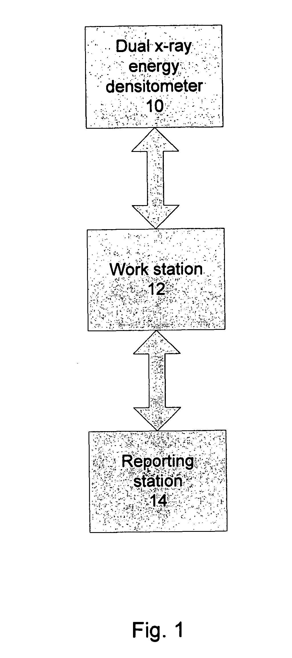
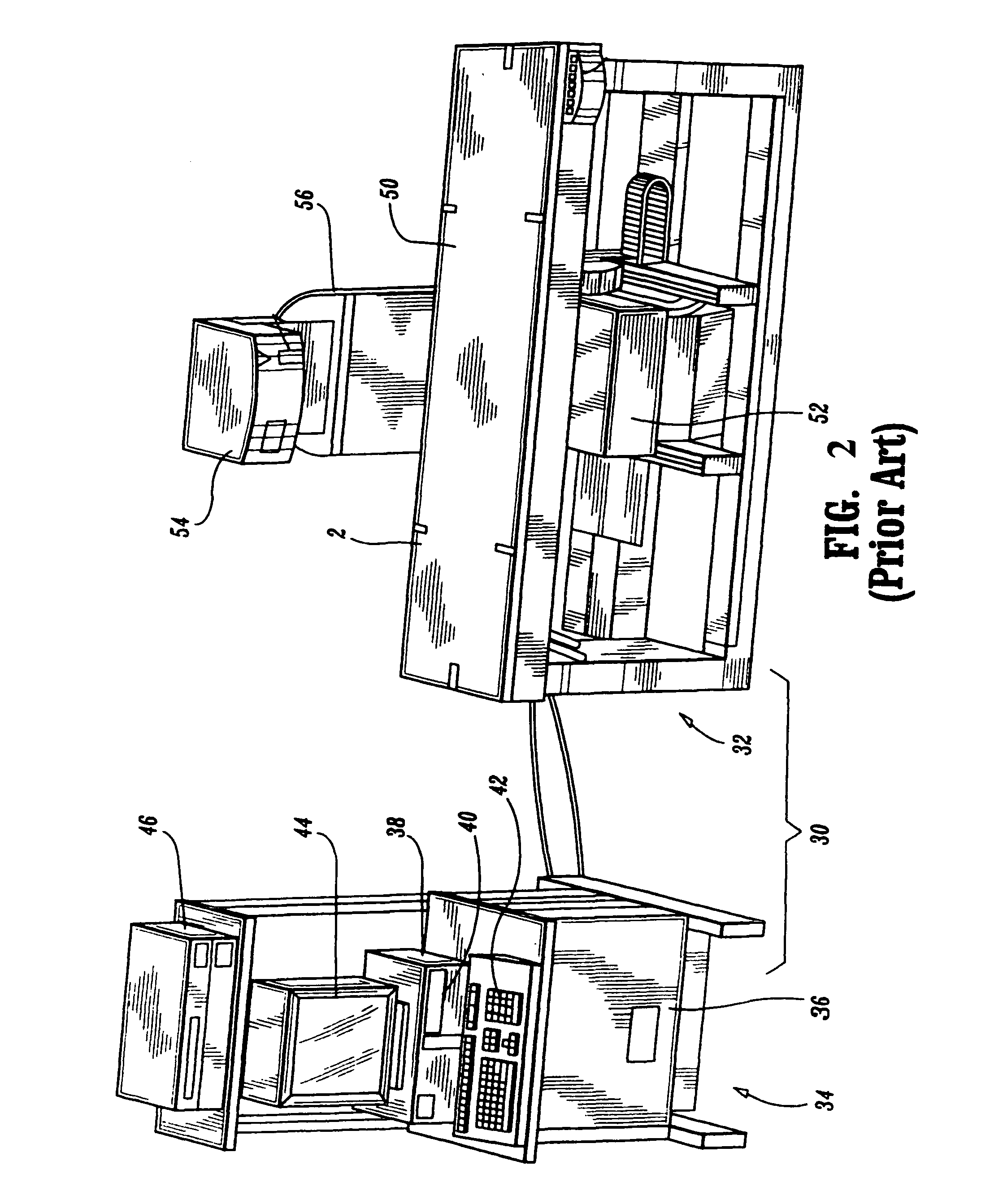
[0015]Referring to FIG. 1, the main components of a system carrying out one example of the disclosed method of estimating and reporting a risk of a cardiovascular event are a dual x-ray energy bone densitometer 10, a processing workstation 12, and a reporting station 14. Densitometer 10 can be the scanning and pre-processing part of a device such as disclosed in U.S. Pat. No. 6,385,283, which is hereby incorporated by reference, or the densitometer available from Hologic, Inc. under the trade name Discovery and equipped with appropriate software including IVA software. Its purpose here is to obtain x-ray measurements of patient's anatomy that includes the appropriate part of the aorta, for example the portion of the abdominal aorta anterior to the lumbar spine. While information for similar processing can be derived from conventional radiography systems, or CT systems, or ECT systems, it is believed that using dual x-ray densitometry is particularly advantageous at least because it ...


PUM


Abstract

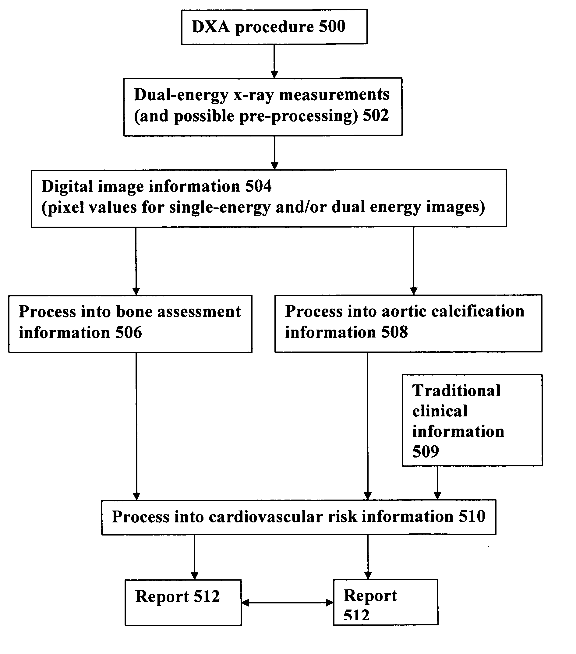
Methods and systems for computer assisted detection of arterial calcification, for example in the abdominal artery, by using measurements such as those conventionally taken with a dual x-ray energy bone densitometers at single energy or dual energy, and for using the calcification assessment either alone or with other information to assess and report a risk of a cardiovascular event.

Description

FIELD AND BACKGROUND[0001]This patent specification is in the field of methods and equipment for assisting medical professionals in assessing cardiovascular risk of patients and bone condition and fracture risk of patients. More particularly, this patent specification pertains to taking x-ray measurements with a dual energy bone densitometer for at least one of the two x-ray energy ranges that the densitometer can measure, and processing these measurements to detect and quantify aortic calcification, to use the results in estimating cardiovascular risk, and to report the outcome in particularly useful ways.[0002]The abdominal aorta is a large, fluid filled vessel comprised of soft tissue. It can be barely visible on conventional x-ray images and is typically is visualized with the use of a radio-opaque contrast material. A vessel that is calcified usually presents with a linear or eggshell calcified pattern which is typical of calcification in the wall of a fluid filled structure. T...

Claims


Application Information

Patent Timeline
Patent Type & Authority Patents(United States)
IPC IPC(8): G06K9/00
CPCA61B5/02007A61B5/7275A61B6/482A61B6/505G06F19/345G06F19/3487G06T7/0012G06T2207/10116G06T2207/30012G06T2207/30048G06T2207/30101G16H15/00G16H50/20Y10S128/922
Inventor WILSON, KEVINHERRINGTON, BRAD
Owner HOLOGIC INC
Learn More