Methods for inhibiting angiogenesis

an angiogenesis and angiogenesis technology, applied in the direction of angiogenin, drug compositions, peptides, etc., can solve the problems of inability to detect and inhibit angiogenesis, and low survival rate of most cancers, so as to inhibit angiogenesis, inhibit angiogenesis in skin tissue, and inhibit angiogenesis.

Inactive Publication Date: 2010-11-02
RGT UNIV OF CALIFORNIA
View PDF21 Cites 5 Cited by
  • Summary
  • Abstract
  • Description
  • Claims
  • Application Information

AI Technical Summary

Problems solved by technology

However, inappropriate angiogenesis can have severe consequences.
Although advances in therapy and in our understanding of cancer causes and risk factors have lead to improved outcomes overall, most cancers still have low five year survival rates.
For example, chemotherapeutics reagents or radiation have serious side effects because they kill or impair all proliferating cells in the body, including healthy cells.
Side effects are unpleasant and often create health problems that themselves increase patient mortality.
In addition, the growth of capillaries into atherosclerotic plaques is a serious problem; the rupture and hemorrhage of vascularized plaques is thought to cause coronary thrombosis.
To date, however, no effective therapies exist for these diseases.
Vascularization in ocular tissue has adverse effects on vision.
New blood vessels on the cornea can induce corneal scarring, whereas new blood vessels on the retina can induce retinal detachment, and angiogenic vessels in the choroid may leak vision-obscuring fluids; these events often lead to blindness.
For other pathological conditions associated with abnormal angiogenesis such as diabetic retinopathy, there are no effective treatments short of retinal transplants.
However, even in cases where retinal transplantation is performed, the new retina is subject to the same conditions that resulted in the original retinopathy.

Method used


Image

  • Methods for inhibiting angiogenesis
  • Methods for inhibiting angiogenesis
  • Methods for inhibiting angiogenesis

Examples


example 1

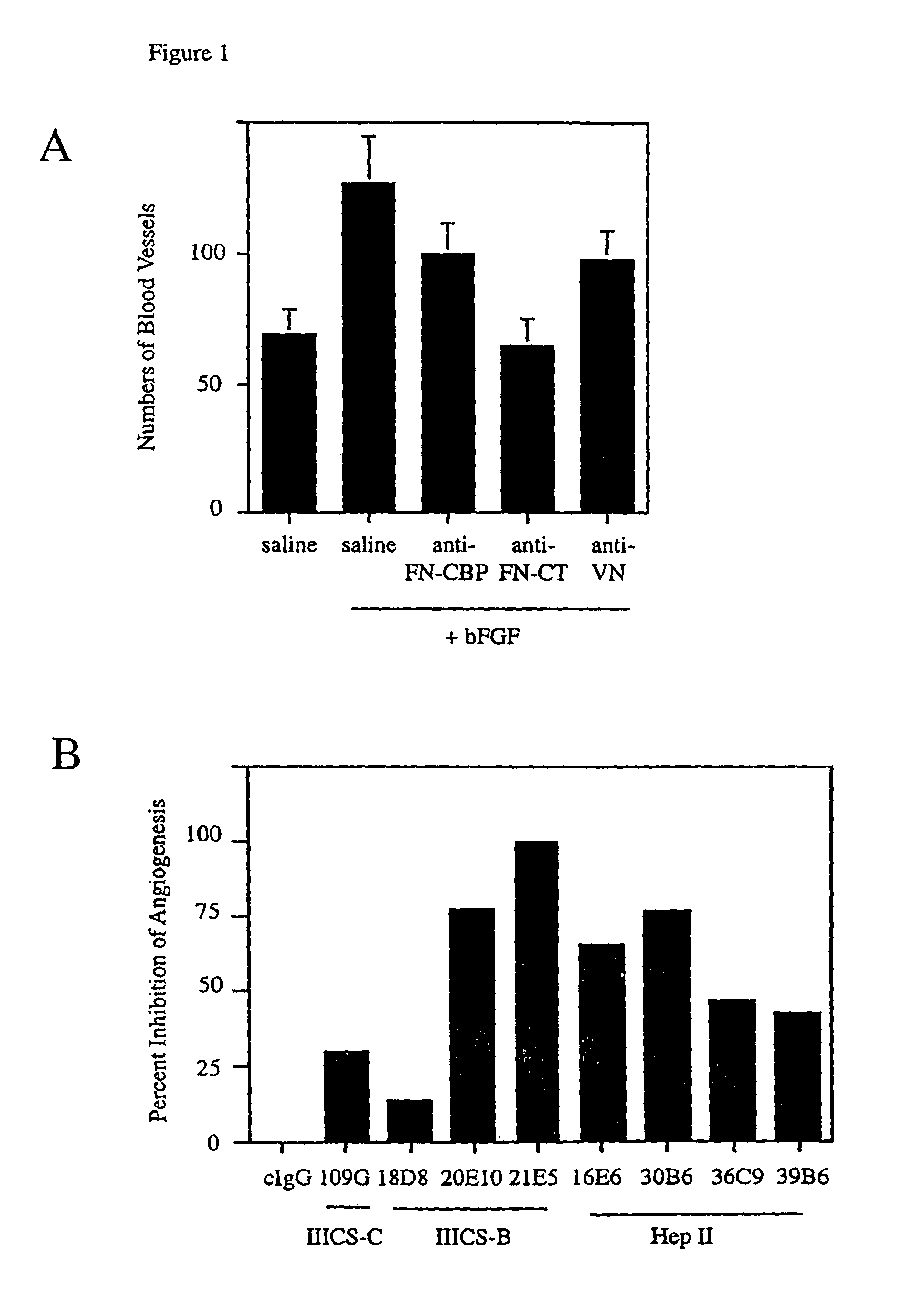
Inhibition of Angiogenesis by Anti-Fibronectin Antibody

[0193]Angiogenesis was stimulated on the surface of the chick chorioallantoic membrane (“CAM”) by applying a 5 mm filter disk on the CAM saturated with basic fibroblast growth factor (“bFGF”), vascular endothelial growth factor (“VEGF”), interleukin 8 (“IL-8”), tumor necrosis factor alpha (“TNF-α”) or saline. Twenty four hours after angiogenesis had been stimulated saline, 25 μg control IgG, 25 μg of an antibody directed against the cell binding peptide of fibronectin (Anti-CPB), 25 μg of an antibody directed against the fibronectin C-terminus (Anti-CT), 25 lag of an antibody directed against the cell binding peptide of fibronectin (Anti-CBP) or 25 μg of an antibody directed against vitronectin (Anti-VN) were applied to the CAM of eight to ten eggs each in a volume of 25 μl. Forty eight hours after this, CAMs were excised from the egg and the number of blood vessels in each filter disk were counted. The results are shown in FIG....

example 2

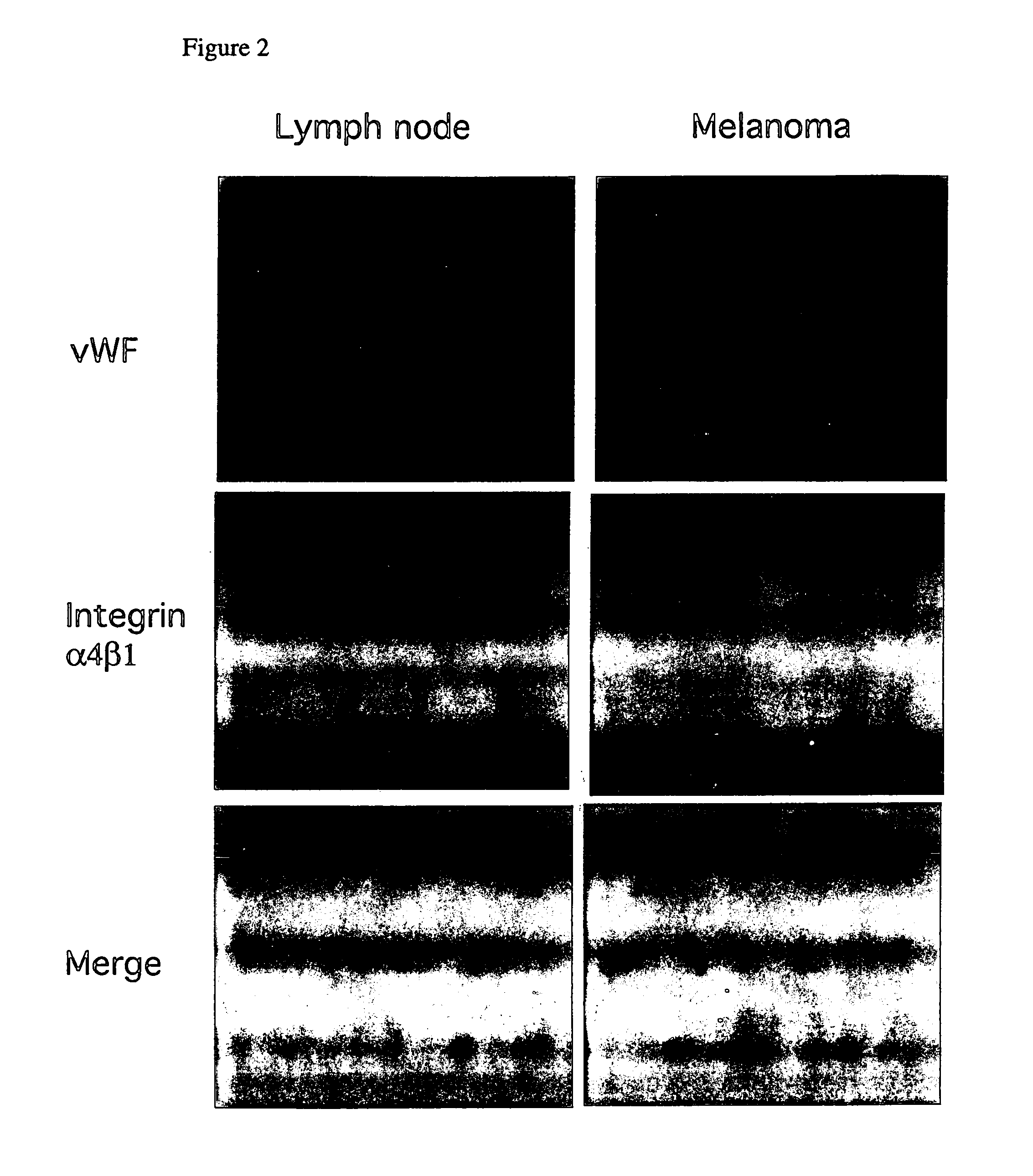
Immunohistochemical Analysis of Integrin α4β1 Expression in Normal and Tumor Tissue

[0196]Five micron frozen sections of human normal thymus (FIG. 2A) and melanoma (FIG. 2B) were fixed for 2 minutes in acetone, air dried and rehydrated for 5 minutes in phosphate buffered saline. Sections were then blocked for 2 hours in a 2% bovine serum albumin in phosphate buffered saline and incubated with 5 μg / ml anti-α4β1 monoclonal antibody and anti-von Willebrand Factor (a marker of endothelial cells used by pathologists to identify blood vessels) polyclonal antibody for one hour room temperature. Sections were washed in PBS and incubated in 1:400 dilution of goat anti-rabbit-FITC and in goat anti-mouse-rhodamine for 1 hour at room temperature. Slides were well washed, and coverslips were mounted in one drop of Fluoromount, prior to photography under fluorescent illumination. Blood vessels positive vWF (von Willebrand Factor) only are green, cells positive for integrin α4β1 only are red and bl...

example 3

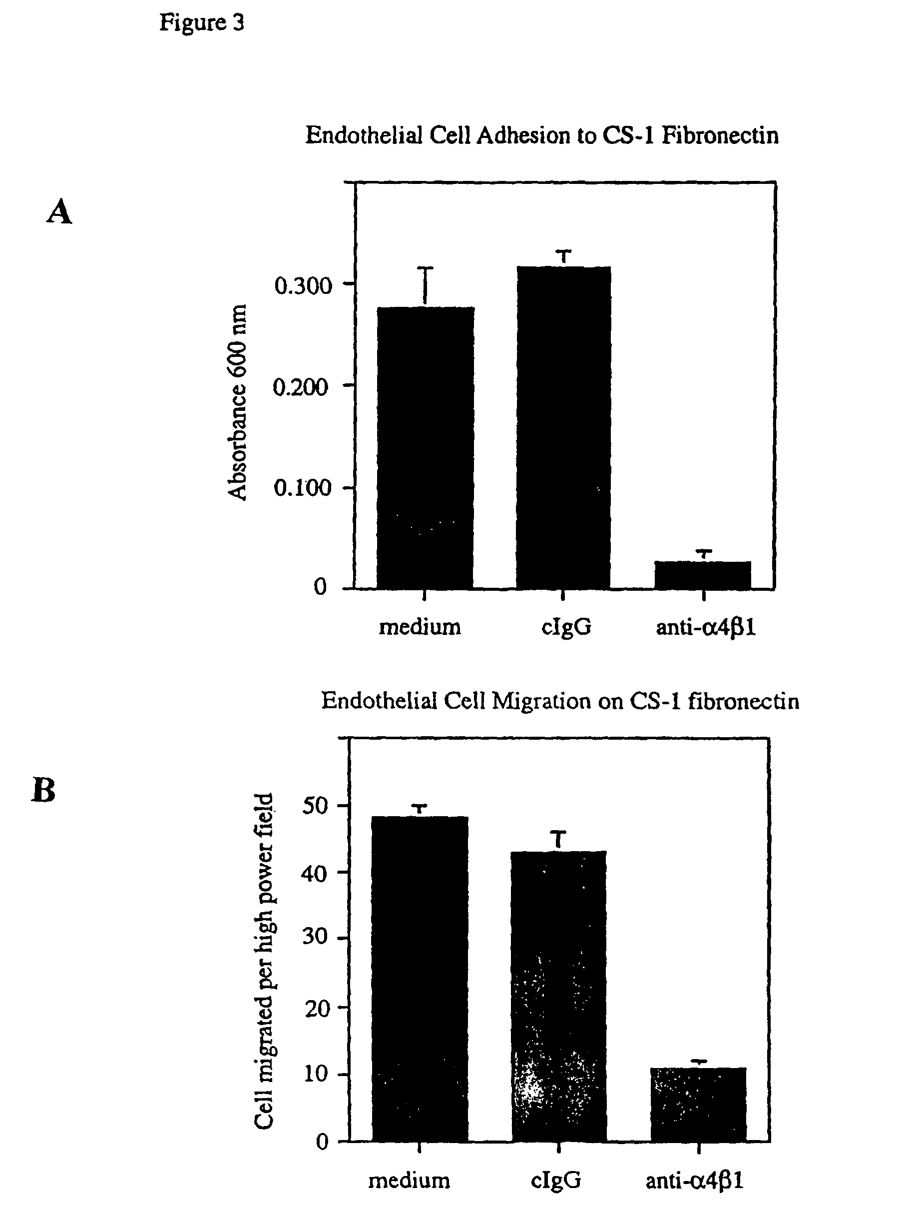
Inhibition of Human Neonatal Cell Adhesion to, and Migration of Human Vascular Endothelial Cells on, CS-1 Fibronectin by Anti-Integrin α4β1 Antibody

1. Endothelial Cell Adhesion:

[0198]The wells of 48 well culture dishes were coated with 10 μg / ml CS-1 fibronectin for one hour at 37° C. and blocked with 2% bovine serum albumin in phosphate buffered saline for one hour. Fifty thousand cells in 250 μl of adhesion buffer were added in triplicate to wells containing 250 μl of a solution of 50 μg / ml of an anti-α4β1 blocking antibody in adhesion buffer, 50 μg / ml of control antibody in adhesion buffer or adhesion buffer (Hepes buffered Hanks balanced salt solution, HBSS). Human neonatal umbilical vein endothelial cells were allowed to adhere to dishes for ten to twenty minutes at 37° C. Nonadherent cells were removed by washing each well four times with 500 μl of adhesion buffer. Adherent cells were then fixed for 15 minutes with 3.7% paraformaldehyde in phosphate buffered saline and stained ...


PUM

PropertyMeasurementUnit
volumeaaaaaaaaaa
anti-vascular cell adhesionaaaaaaaaaa
vascular cell adhesionaaaaaaaaaa

Abstract

The invention provides methods for detecting and inhibiting angiogenesis, endothelial cell adhesion, and endothelial cell migration using agents which inhibit the specific binding of integrin α4β1 to one or more of its ligands. The invention further provides methods for screening test compounds for their ability to inhibit angiogenesis, endothelial cell adhesion, or endothelial cell migration by employing agents which inhibit the specific binding of integrin α4β1 to one or more of its ligands. The invention additionally relates to methods for isolating endothelial progenitor cells which express integrin α4β1. The methods of the invention are useful in, for example, diagnosing and inhibiting pathological conditions that are associated with angiogenesis, endothelial cell adhesion, and / or endothelial cell migration. The invention's methods are also useful in isolating endothelial progenitor cells, and in determining the mechanisms that underlie angiogenesis, development, wound healing, and the function of the female reproductive system.

Description

[0001]This application is the U.S. National stage filing of PCT Application No. PCT / US02 / 24573, filed on Aug. 1, 2002, which claims priority to U.S. provisional Patent Application Ser. No. 60 / 310,645, filed Aug. 6, 2001, now abandoned.[0002]This invention was made, in part, with government support under grant numbers CA71619 and CA 83133 awarded by the National Cancer Institute of the National Institutes of Health. The U.S. government has certain rights in the invention.FIELD OF THE INVENTION[0003]The present invention provides methods for detecting and inhibiting angiogenesis, endothelial cell adhesion, and endothelial cell migration. In preferred embodiments, the present invention utilizes agents that inhibit the specific binding of integrin α4β1 to one or more of its ligands. The invention further relates to methods for screening test compounds for their ability to inhibit angiogenesis, endothelial cell adhesion, or endothelial cell migration by employing agents which inhibit the...

Claims


Application Information

Patent Timeline
Patent Type & Authority Patents(United States)
IPC IPC(8): A61K39/395A61K38/10A61K38/39A61K48/00C07K16/18C07K16/28G01N33/50G01N33/68
CPCA61K38/39C07K16/18C07K16/2842G01N33/5011G01N33/6893A61K2039/505C07K2316/96G01N2333/70546G01N2333/7055G01N2500/04G01N2500/10A61P35/00C07K2317/76
Inventor VARNER, JUDITH A.
Owner RGT UNIV OF CALIFORNIA
Why Eureka
Try Eureka