Microfluidic chip and method for studying non-contact type cell co-cultivation by using the same

A microfluidic chip and cell culture technology, applied in the field of biomedical research, can solve the problems of large consumption of reagents, lack of biological information, and large amount of cells, and achieve the effects of less sample consumption, simple production and simple operation.

Inactive Publication Date: 2011-04-20
DALIAN INST OF CHEM PHYSICS CHINESE ACAD OF SCI
View PDF1 Cites 28 Cited by
  • Summary
  • Abstract
  • Description
  • Claims
  • Application Information

AI Technical Summary

Problems solved by technology

Mixed co-cultivation, two or more types of cells cannot be distinguished; microcarrier co-cultivation, cell interaction time is limited, generally less than 4 hours; and for co-cultivation methods such as Transwell, it is impossible to monitor in real time Cellu

Method used


Image

  • Microfluidic chip and method for studying non-contact type cell co-cultivation by using the same
  • Microfluidic chip and method for studying non-contact type cell co-cultivation by using the same
  • Microfluidic chip and method for studying non-contact type cell co-cultivation by using the same

Examples


Embodiment 1

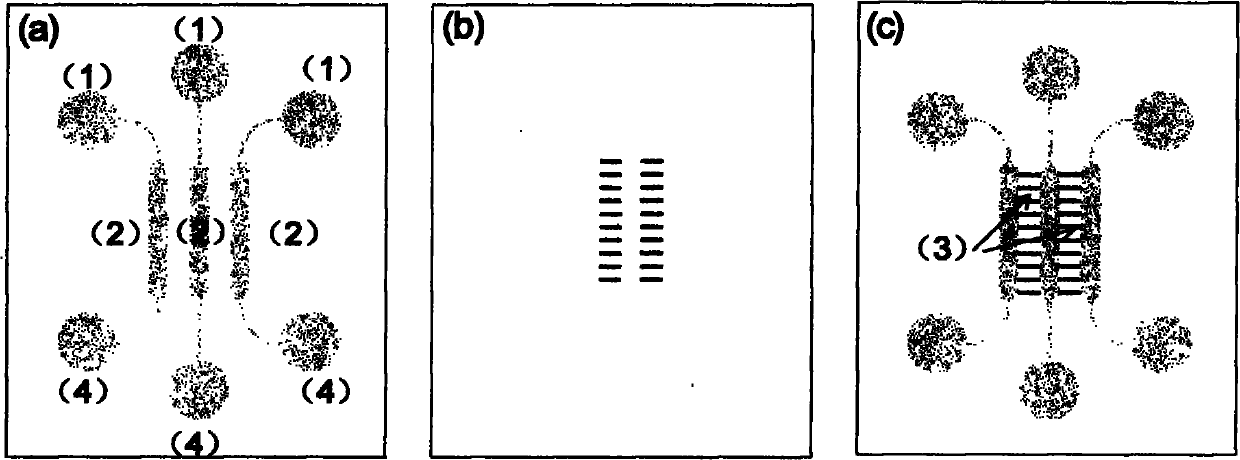
[0030] The microfluidic chip used was designed and manufactured by our laboratory. The chip is formed by reversible sealing of upper and lower layers, the upper layer material is PDMS polymer, and the lower layer material is glass. like figure 1 As shown, the dimensions of three parallel cell culture chambers are 6 mm in length, 1 mm in width, and 50 μm in height, and the dimensions of the cell migration area are 1 mm in length, 500 μm in width, and 50 μm in height. Add hepatoma cell HepG-2 into cell culture chamber C through the injection port on the right, and gently place it in 37°C CO 2 In the incubator; after the cells adhere to the wall, add the fibroblast HFL-I into the cell culture chamber B through the middle inlet; as a blank control, the left culture chamber A does not add cells, and adds the same amount of cell culture medium as B and C .

[0031] Hoechst33342 and PI were used to jointly detect the activity of three kinds of cells (HepG-2, GES-1, HFL-I), and the...

Embodiment 2

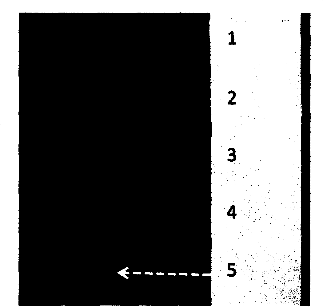
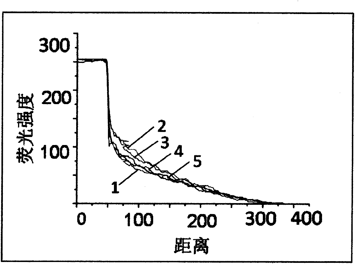
[0034] Embed fluorescent dye labeled with fluorescein isothiocyanate (FITC) in BME, add it into cell culture chamber C through the injection port, and add an equal amount of α-MEM culture solution to the rest of the cell culture chambers. After a period of time, the cells A fluorescent signal appeared in the migration area ( figure 2 ), data analysis shows that the cell migration area forms a stable fluorescence concentration gradient ( image 3 ).

[0035] Add the normal cell GES-1 into the cell culture chamber C through the injection port on the right, and gently place it in the CO at 37°C. 2 In the incubator; after the cells adhere to the wall, add the fibroblast HFL-I into the cell culture chamber B through the middle inlet; as a blank control, the left culture chamber A does not add cells, and adds the same amount of cell culture medium as B and C . Compared with the migration of HFL-I cells on the blank control side, at 6h and 48h, the migration number and area of ​​...

Embodiment 3

[0037] Add oral cancer cells ACC-2 / ACC-M into cell culture chamber C through the injection port on the right, and gently place it in 37°C CO 2 In the incubator; after the cells adhere to the wall, add the fibroblast HFL-I into the cell culture chamber B through the middle inlet; as a blank control, the left culture chamber A does not add cells, and adds the same amount of cell culture medium as B and C . Compared with the HFL-I cell migration on the blank control side, there was no significant difference in the HFL-I cell migration area on the experimental side and the control side under the induction of oral cancer cell ACC-2 / ACC-M ( Figure 8 , Figure 9 ).


PUM


Abstract

The present invention provides a microfluidic chip and a method for studying non-contact type cell co-cultivation by using the same. The microfluidic chip comprises a sample inlet, a cell cultivation room, a cell migration region and a waste pool. The upper end of the cell cultivation room is connected to the sample inlet and the lower end of the cell cultivation room is connected to the waste pool. Three parallel cell cultivation rooms are joined by cell migration region. The microfluidic chip provides good microenvironment for non-contact type cell co-cultivation, enables to form a concentration gradient in the cell migration region and facilitates observation of cell migration process in real time and research of transdifferentiation. The invention is advantageous in easy operation, simple manufacturing and small dosage of samples.

Description

technical field [0001] The invention relates to the application of microfluidic chip technology to the field of biomedical research, and specifically provides a microfluidic chip and a method for researching non-contact cell co-cultivation. Background technique [0002] Cell interaction is a research hotspot in the fields of medicine and biology today. A cell is not an isolated unit of life, nor is there a simple mechanical connection between cells, but there are various forms of mutual communication to form a complete individual and coordinate various life activities. Exploring the interaction between cells and the corresponding signaling pathways plays an important role in the treatment of diseases. [0003] Currently, cell co-culture is the most commonly used method to study cell interactions. Traditionally, cell co-culture methods are generally divided into three categories: mixed co-culture, microcarrier co-culture and Transwell co-culture. Mixed co-cultivation, two ...

Claims


Application Information

Patent Timeline
IPC IPC(8): C12M3/00C12Q1/02
Inventor 秦建华马慧朋林炳承
Owner DALIAN INST OF CHEM PHYSICS CHINESE ACAD OF SCI
Why Eureka
Try Eureka