Derivation of embryonic stem cells and embryo-derived cells

a technology of embryonic stem cells and embryos, which is applied in the field of embryonic stem cells and embryo-derived cells, can solve the problems of es cell-stage blastocyst embryos that cannot be cryopreserved, frozen for later use, or allowed to develop further

Active Publication Date: 2011-02-22
ADVANCED CELL TECH INC
View PDF20 Cites 36 Cited by
  • Summary
  • Abstract
  • Description
  • Claims
  • Application Information

AI Technical Summary

Problems solved by technology

There are several drawbacks to the techniques used to create these cells.
From the perspective of the technique, the culturing of embryos to blastocysts occasionally has a relatively low success rate.
Additionally, certain members of the public object to embryonic stem (ES) cell research using cell lines derived from the inner cell mass of blastocysts because this derivation procedure destroys the preimplantation, blastocyst-stage embryo.
As such, the blastocyst-stage embryo from which ES cells are conventionally produced cannot be cryopreserved, frozen for later use, or permitted to develop further.

Method used


Image

  • Derivation of embryonic stem cells and embryo-derived cells
  • Derivation of embryonic stem cells and embryo-derived cells
  • Derivation of embryonic stem cells and embryo-derived cells

Examples


example 1

Generation of Human ES Cell Lines

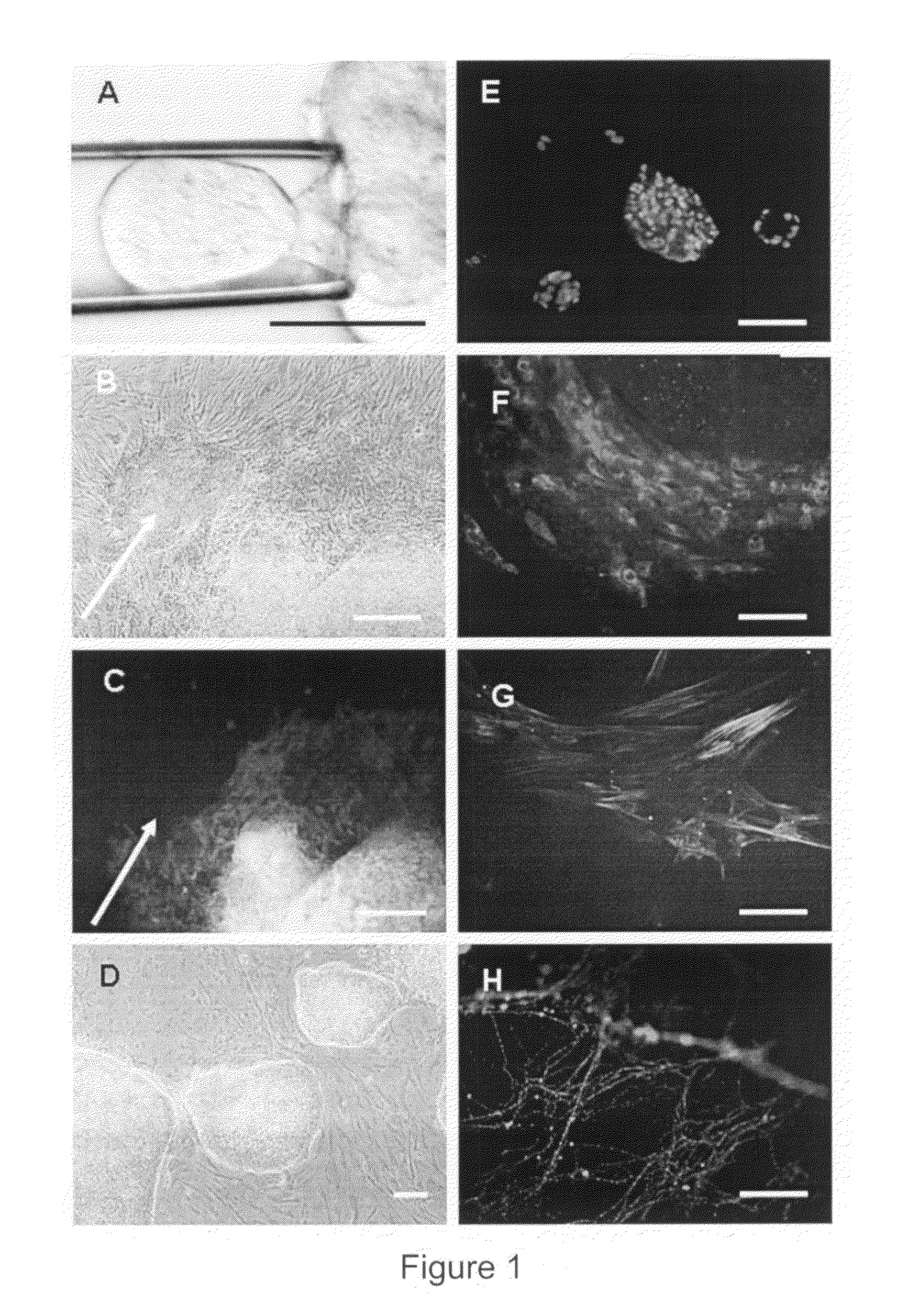
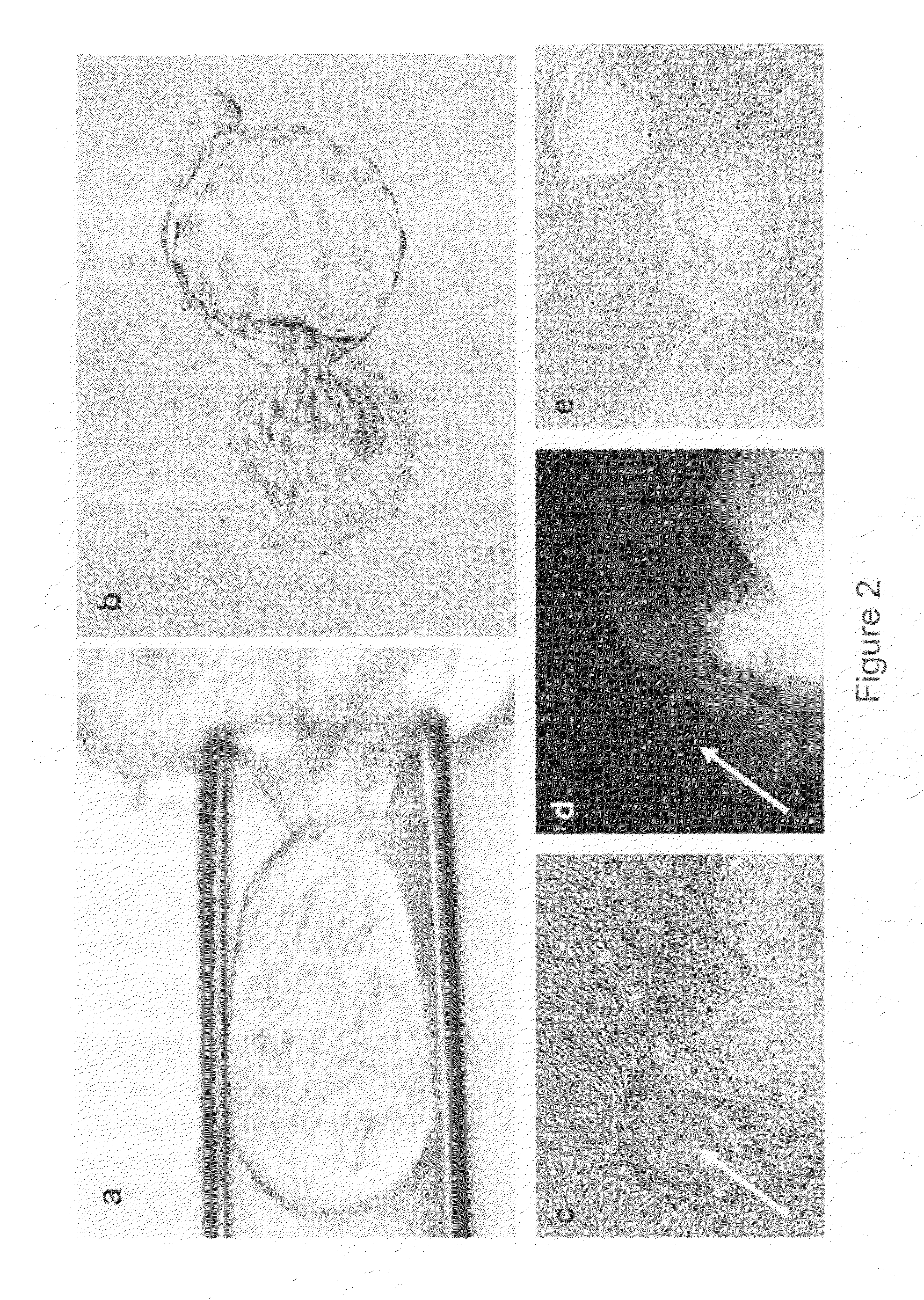
Unused embryos produced by in-vitro fertilization for clinical purposes were obtained. Six of these embryos were Grade I or II (symmetrical and even cell division with little or no cytoplasmic fragmentation), whereas the remaining ten embryos were Grade III (variable fragmentation) using standard scoring system (Veeck, L. L. et al., An Atlas of Human Gametes and Conceptuses, Parthenon, New York, N.Y., 1999). Embryos with blastomeres of unequal size and moderate-to-severe fragmentation (Grades IV and V) were excluded from this study. Pronuclear and multi-cell stage human embryos were thawed and cultured until the 8-10 cell stage at 37 C in 20 μl drops of Quinn's cleavage medium (Cooper Surgical Inc., Cat # ART1526) under paraffin oil (Cooper Surgical Inc. Cat # 4008) in a high humidified incubator with 5.5% CO2 / 5% O2 / 89.5% N2.

The zona pellucida was disrupted using either Acidic Tyroides solution or multiple Piezo-pulses and individual blastomeres were...

example 2

Differentiation of Human ES Cells

The ability of the human ES cells to differentiate into different germ layers was analyzed both in vitro and in teratomas using techniques known in the art.

Briefly, for the in vitro experiments, the human ES cells were separated by treating with either collagenase or trypsin and then cultured in cell culture dishes without feeder cells in embryoid body (EB) medium. Approximately one week later, the ES cells formed embryoid bodies (EB). The EBs were then fixed in 4% formaldehyde, washed in PBS, embedded in paraffin, sectioned and analyzed for the presence of derivatives from endoderm, mesoderm and ectoderm using tissue specific antibodies (α-feto protein for primitive endoderm, muscle actin for mesoderm, and β III tubulin for ectoderm) (FIG. 4).

The single blastomere-derived human ES cell could also be differentiated in vitro into cells of specific therapeutic interest, including endothelial cells which after replating on Matrigel, formed typical capil...

example 3

Production of ED Cells

As can be seen in Table 1 above, the production of embryo-derived cells from isolated blastomeres occurs more often than the production of ES cell lines. Of 53 isolated blastomeres that divided, 19 cultures yielded directly-differentiated cell types and only 2 yielded ES cell lines. FIG. 6 shows the variety of differentiated cell morphologies observed by direct differentiation.


PUM

PropertyMeasurementUnit
osmolarityaaaaaaaaaa
volumeaaaaaaaaaa
osmolarityaaaaaaaaaa

Abstract

This present invention provides novel methods for deriving embryonic stem cells and embryo-derived cells from an embryo without requiring destruction of the embryo. The invention further provides cells and cell lines derived without embryo destruction, and the use of the cells for therapeutic and research purposes. It also relates to novel methods of establishing and storing an autologous stem cell line prior to implantation of an embryo, e.g., in conjunction with reproductive therapies such as IVF.

Description

FIELD OF THE INVENTIONThis invention generally relates to novel methods for deriving embryonic stem (ES) cells and embryo-derived (ED) cells, those cells and cell lines, and the use of the cells for therapeutic and research purposes. It also relates to novel methods of establishing and storing an autologous stem cell line prior to implantation of an embryo, e.g. in conjunction with assisted reproductive technologies such as in vitro fertilization.BACKGROUND OF THE INVENTIONWith few exceptions, embryonic stem cells have only been grown from blastocyst-stage embryos. ES cell lines are conventionally isolated from the inner cell mass of blastocysts. There are several drawbacks to the techniques used to create these cells. From the perspective of the technique, the culturing of embryos to blastocysts occasionally has a relatively low success rate. Additionally, certain members of the public object to embryonic stem (ES) cell research using cell lines derived from the inner cell mass of ...

Claims


Application Information

Patent Timeline
Patent Type & Authority Patents(United States)
IPC IPC(8): A61K35/14A61K35/12C12N5/0735
CPCC12N5/0604C12N5/0606C12N15/873A61K35/12C12N2506/02C12N2502/02C12N2500/34C12N2501/998C12N2501/115A61P43/00C12N5/0603
Inventor CHUNG, YOUNG GIELANZA, ROBERTKLIMANSKAYA, IRINA V.
Owner ADVANCED CELL TECH INC
Why Eureka
Try Eureka