MicroRNA fingerprints during human megakaryocytopoiesis
a microrna fingerprint and megakaryocytic technology, applied in the field of microrna fingerprints during human megakaryocytic differentiation, can solve the problems of patient running the risk of catastrophic hemorrhage, and achieve the effects of determining and/or predicting megakaryocytic differentiation, reducing expression, and reducing expression
- Summary
- Abstract
- Description
- Claims
- Application Information
AI Technical Summary
Benefits of technology
Problems solved by technology
Method used
Image
Examples
example 1
miRNA Expression During In Vitro Megakaryocytic Differentiation of CD34+ Progenitors
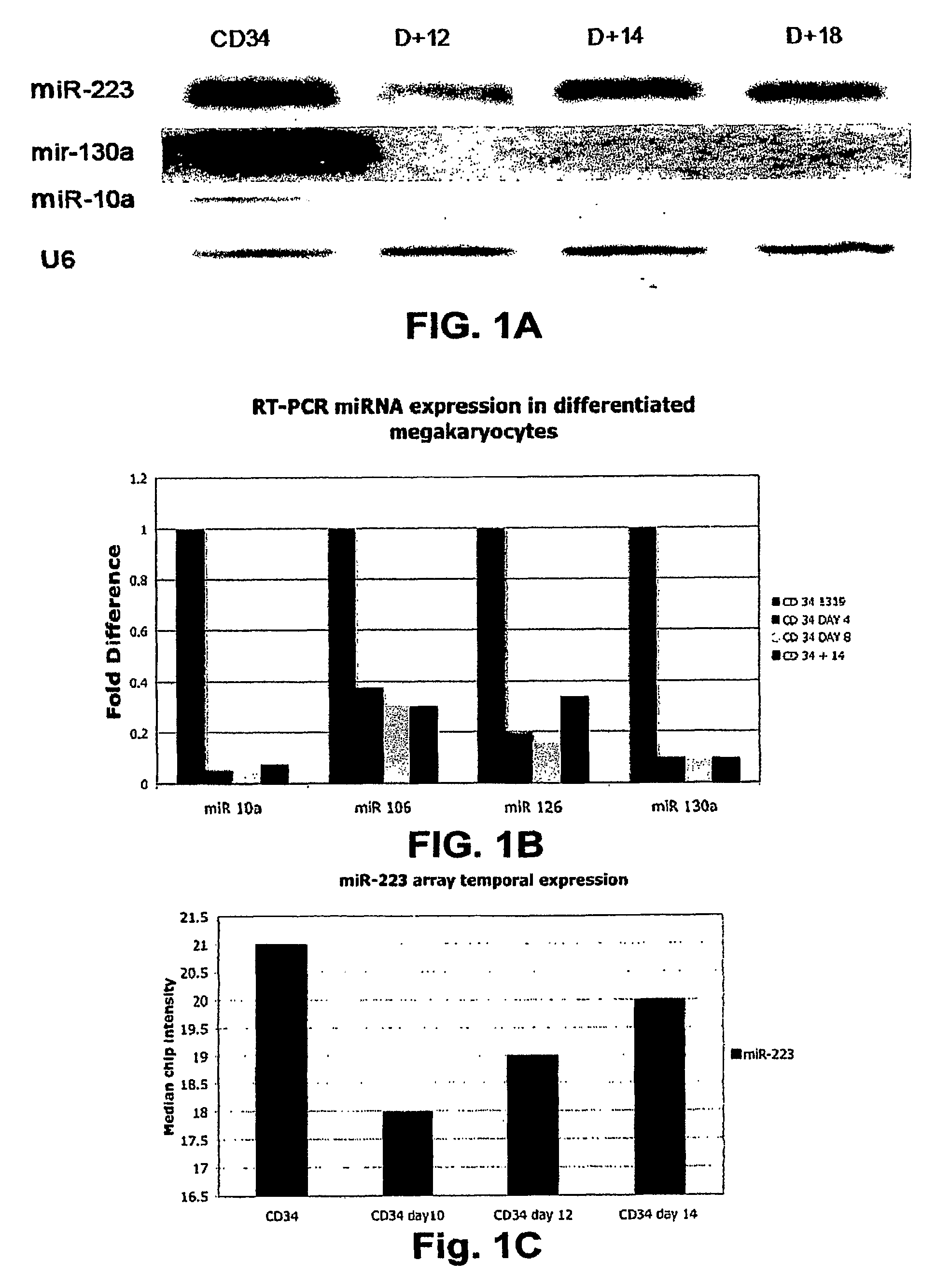
[0195]Using a combination of a specific megakaryocytic growth factor (thrombopoietin) and nonspecific cytokines (SCF and IL-3), we were able to generate in vitro pure, abundant megakaryocyte progeny from CD34+ bone marrow progenitors suitable for microarray studies (FIG. 4). Total RNA was obtained for miRNA chip analysis from three different CD34 progenitors at baseline and at days 10, 12, 14 and 16 of culture with cytokines. We initially compared the expression of miRNA between the CD34+ progenitors and the pooled CD34+ differentiated megakaryocytes at all points during the differentiation process. 17 miRNAs (Table 1) that are sharply down regulated during megakaryocytic differentiation were identified. There were no statistically significant miRNAs upregulated during megakaryocytic differentiation. Using predictive analysis of microarray (PAM), we identified 8 microRNAs that predicted megakaryocyti...
example 2
MAFB Transcription Factor is a Target of miR-130a
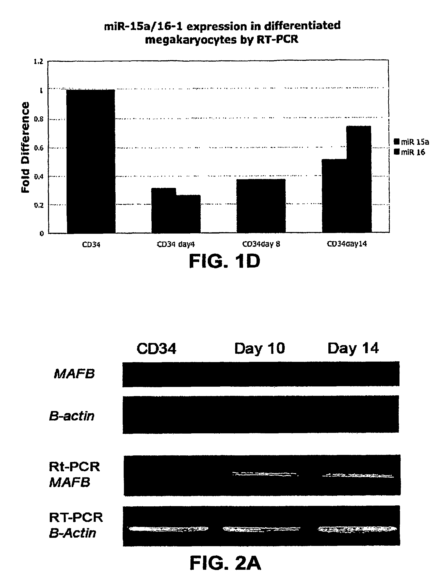
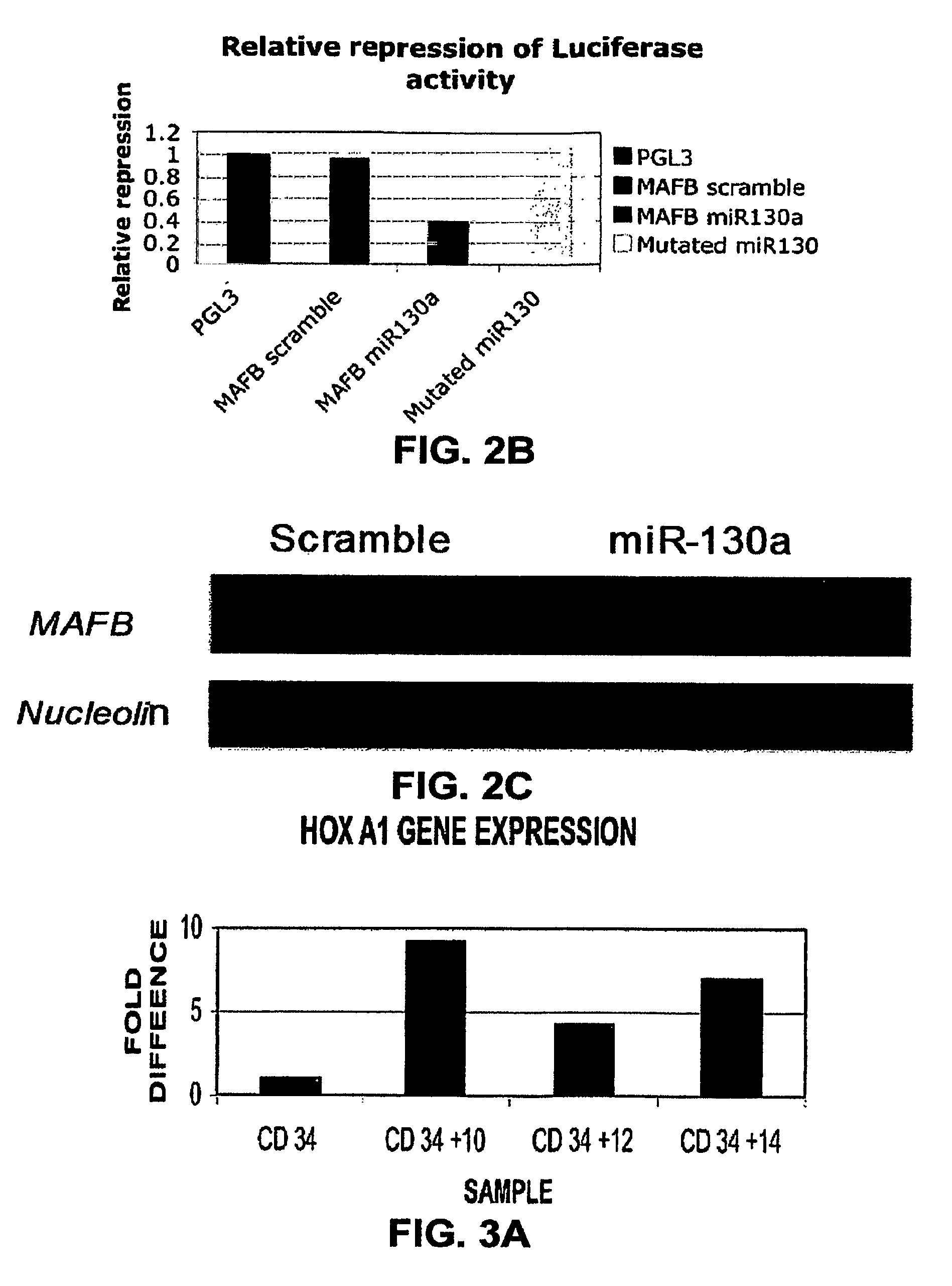
[0200]By using three target prediction algorithms (TARGETSCAN (www.genes.mit.edu / targetscan), MIRANDA (www.microma.org / miranda_new.htm 1), and PICTAR (www.pictar.bio.nyu.edu)), we identified that miR-130a is predicted to target MAFB, a transcription factor that is upregulated during megakaryocytic differentiation and induces the GPIIb gene, in synergy with GATA1, SP1 and ETS-1 (Sevinsky, J. R., et al. (2004) Mol. Cell. Biol. 24, 4534-4545). To investigate this putative interaction, first, we examined MAFB protein and mRNA levels in CD34+ progenitors at baseline and after cytokine stimulation (FIG. 2A). We found that the MAFB protein is upregulated during in vitro megakaryocytic differentiation. Although the mRNA levels for MAFB by PCR increase with differentiation, this increase does not correlate well with the intensity of its protein expression. The inverse pattern of expression of MAFB and miR-130a suggested in vivo interaction tha...
example 3
MiR-10a Correlates with HOXB Gene Expression
[0203]It has been reported that in mouse embryos, miR-10a, miR-10b, and miR-196 are expressed in HOX-like patterns (Mansfield, J. H., et al. (2004) Nature 36, 1079-1083) and closely follow their “host” HOX cluster during evolution (Tanzer, A., et al. (2005) J. Exp. Zool. B Mol. Dev. Evol. 304B, 75-85). These data suggest common regulatory elements across paralog clusters. MiR-10a is located at chromosome 17q21 within the cluster of the HOXB genes (FIG. 8) and miR-10b is located at chromosome 2q31 within the HOXD gene cluster. To determine whether the miR-10a expression pattern correlates with the expression of HOXB genes, we performed RT-PCR for HOXB4 and HOXB5, which are the genes located 5′ and 3′, respectively, to miR-10a in the HOXB cluster. As shown in FIG. 8, HOXB4 and HOXB5 expression paralleled that of miR-10a, suggesting a common regulatory mechanism.
PUM
| Property | Measurement | Unit |
|---|---|---|
| weight | aaaaa | aaaaa |
| volume | aaaaa | aaaaa |
| number-average molecular weight | aaaaa | aaaaa |
Abstract
Description
Claims
Application Information
 - R&D
- Intellectual Property
- Life Sciences
- Materials
- Tech Scout
- Unparalleled Data Quality
- Higher Quality Content
- 60% Fewer Hallucinations
Browse by: Latest US Patents, China's latest patents, Technical Efficacy Thesaurus, Application Domain, Technology Topic, Popular Technical Reports.
© 2025 PatSnap. All rights reserved.Legal|Privacy policy|Modern Slavery Act Transparency Statement|Sitemap|About US| Contact US: help@patsnap.com


