Methods for detecting and treating autoimmune disorders

a technology for autoimmune disorders and autoimmune diseases, applied in the field of autoimmune disorders, can solve the problems of affecting the normal affecting the function of the immune system, and affecting the ability of the immune system to adapt to the expansion of the body, so as to inhibit the autoimmune disease and increase the expression of foxp3

Active Publication Date: 2011-11-08
OREGON HEALTH & SCI UNIV +2
View PDF38 Cites 6 Cited by
  • Summary
  • Abstract
  • Description
  • Claims
  • Application Information

AI Technical Summary

Benefits of technology

[0010]Disclosed herein is a method for inhibiting an autoimmune disease. The method includes administering to a subject a therapeutically effective amount of a composition that increases FOXP3 expression, thereby inhibiting the autoimmune disease.

Problems solved by technology

Autoimmune diseases such as multiple sclerosis (MS) may result from the failure of tolerance mechanisms to prevent expansion of pathogenic T cells directed at myelin determinants or other self-tissue antigens.
Multiple sclerosis can cause blurred vision, unilateral vision loss (optic neuritis), loss of balance, poor coordination, slurred speech, tremors, numbness, extreme fatigue, changes in intellectual function (such as memory and concentration), muscular weakness, paresthesias, and blindness.
Many subjects develop chronic progressive disabilities, but long periods of clinical stability may interrupt periods of deterioration.
Because it is not contagious, which would require U.S. physicians to report new cases, and because symptoms can be difficult to detect, the incidence of disease is only estimated and the actual number of persons with MS could be much higher.
Demyelination interferes with conduction of nerve impulses, which produces the symptoms of multiple sclerosis.

Method used


Image

  • Methods for detecting and treating autoimmune disorders
  • Methods for detecting and treating autoimmune disorders
  • Methods for detecting and treating autoimmune disorders

Examples


example 1

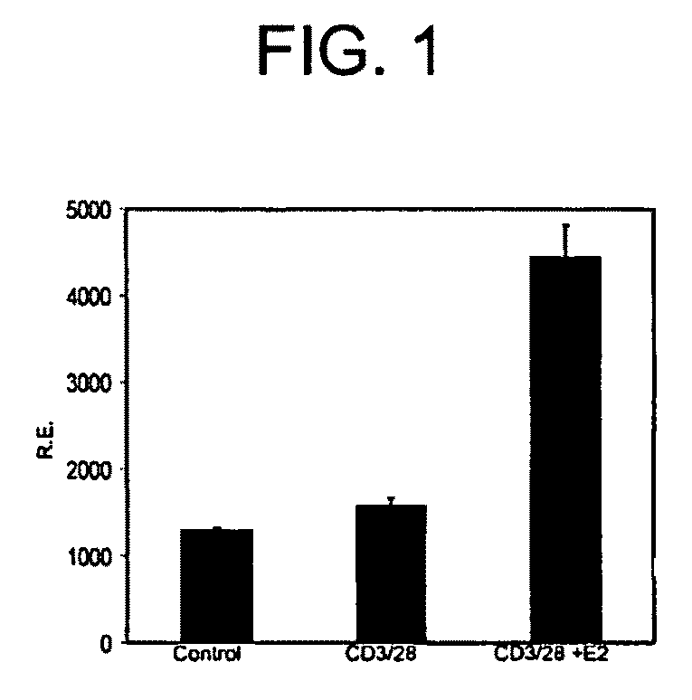
E2 Augments Foxp3 Expression in vitro

[0177]The capacity of E2 to induce Foxp3 expression in vitro in purified (>99%) CD4+CD25− T cells was tested. E2 in combination with TCR stimulation by anti-CD3 antibody for 24 hours induced Foxp3 mRNA approximately 3-fold over levels in untreated cells, while TCR stimulation with anti-CD3 without E2, failed to induce Foxp3 (FIG. 1). In addition, E2 increased the fraction of CD25+ cells over TCR stimulation alone, consistent with induction of Treg.

example 2

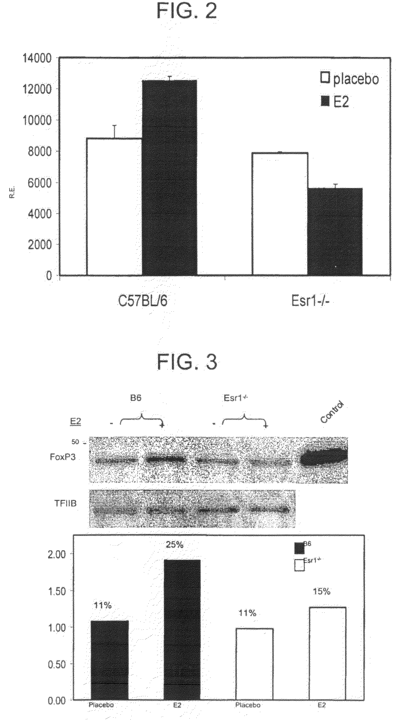
E2 Treatment Before EAE Induction Augments Foxp3 Expression In Vivo

Mice

[0178]Female naïve or syngeneic pregnant (19 days) C57BL / 6 mice were purchased from Jackson Laboratory (Bar Harbor, Me.). Estrogen receptor alpha knockout (Esrl− / −) mice were purchased from Taconic (Germantown, N.Y.). Most experiments represent cells pooled from at least five mice per experimental condition.

Hormone Treatment

[0179]For E2 therapy, a 3 mm pellet containing 2.5 mg or 15 mg (as indicated) 17β-estradiol (Innovative Research of America, Sarasota, Fla.) was implanted s.c. (dorsally) 7 days prior to immunization (EAE) or 14 days prior to analysis of naïve mice. These pellets are designed to release their contents at a constant rate over 60 days. Control animals were implanted with pellets containing saline. Serum levels of E2 were monitored by RIA.

Induction of EAE

[0180]Briefly, mice were immunized s.c. in the flanks with 200 μg MOG-35-55 peptide in CFA (Difco Laboratories, Detroit, Mich.). Mice were also ...

example 3

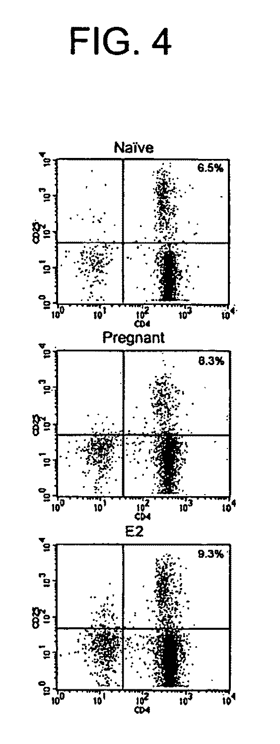
E2 Expands the Treg Compartment in vivo

Cell Preparation and Culture

[0182]Single-cell suspensions were prepared from spleens and RBCs lysed. Non-immunized mice receiving E2 pellets were sacrificed 14 days after implantation, while immunized mice were sacrificed at the peak of EAE disease severity, approximately 17 days after induction. Purified CD4+ cells were obtained by magnetically activated cell sorting (MACS) according to manufacturer's protocols (Miltenyi Biotec, Bergisch Gladbach, Germany). For flow cytometry, cells were stained with FITC-anti-CD4 and PE-anti-CD25 (BD PharMingen, San Diego, Calif.). CD4+CD25− cells were obtained from purified CD4+ using a FACSVantage (BD Immunocytornetry Systems, San Jose, Calif.). For in vitro experiments, CD4+CD25− cells were stimulated with 5 μg / ml anti-CD3ε and 1 μg / ml anti-CD28 (145-2C11 and 37.51, respectively, BD PharMingen). In some experiments, cells were treated with E2 alone in the absence of antibody stimulation.

Evaluation of Foxp3...


PUM

PropertyMeasurementUnit
concentrationaaaaaaaaaa

Abstract

The present disclosure relates to methods for inhibiting an autoimmune disease by administering to a subject a therapeutically effective amount of a composition that increases FOXP3 expression, thereby inhibiting the autoimmune disease. Further disclosed herein are methods for detecting in a subject an autoimmune disease or a predisposition to an autoimmune disease, and methods for assessing the efficacy of a therapy for an autoimmune disease.

Description

CROSS REFERENCE TO RELATED APPLICATIONS[0001]This application is the U.S. National Stage of International Application No. PCT / US2005 / 026915, filed Jul. 29, 2005, which was published in English under PCT Article 21(2), which in turn claims the benefit of U.S. Provisional Patent Application No. 60 / 592,431 filed Jul. 30, 2004 and U.S. Provisional Patent Application No. 60 / 667,820 filed Apr. 1, 2005, both of which are incorporated herein by reference in their entirety.STATEMENT OF GOVERNMENT SUPPORT[0002]This invention was made with United States government support pursuant to grants NS23444, NS45445 and NS23221 from the National Institutes of Health; grants from the Immune Tolerance Network, the National Multiple Sclerosis Society, The Nancy Davis MS Center Without Walls, and the Department of Veterans' Affairs. The United States government has certain rights in the invention.FIELD OF THE DISCLOSURE[0003]This disclosure relates to the field of autoimmune disorders, specifically to the ...

Claims


Application Information

Patent Timeline
Patent Type & Authority Patents(United States)
IPC IPC(8): G01N33/53
CPCA61K39/0008C07K14/7051G01N33/564A61K2039/55566A61K2300/00A61P37/00
Inventor VANDENBARK, ARTHUR A.OFFNER, HALINABARTHOLOMEW, RICHARD
Owner OREGON HEALTH & SCI UNIV
Why Eureka
Try Eureka