Process for examining mineral samples with X-ray microscope and projection systems

a technology of x-ray microscope and projection system, which is applied in the direction of material analysis using wave/particle radiation, instruments, nuclear engineering, etc., can solve the problems of prone to introducing artifacts and inaccurate existing mineral imaging techniques

Active Publication Date: 2011-11-29
CARL ZEISS X RAY MICROSCOPY
View PDF5 Cites 100 Cited by
  • Summary
  • Abstract
  • Description
  • Claims
  • Application Information

AI Technical Summary

Benefits of technology

[0011]The above and other features of the invention including various novel details of construction and combinations of parts, and other advantages, will now be more particularly described with reference to the accompanying drawings and pointed out in the claims. It will be understood

Problems solved by technology

The existing mineral imaging techniques are typica

Method used


Image

  • Process for examining mineral samples with X-ray microscope and projection systems
  • Process for examining mineral samples with X-ray microscope and projection systems
  • Process for examining mineral samples with X-ray microscope and projection systems

Examples


Embodiment Construction

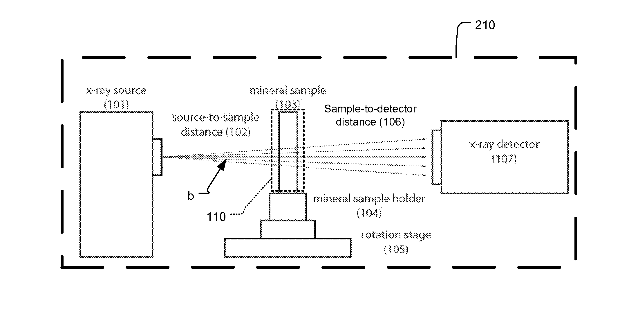
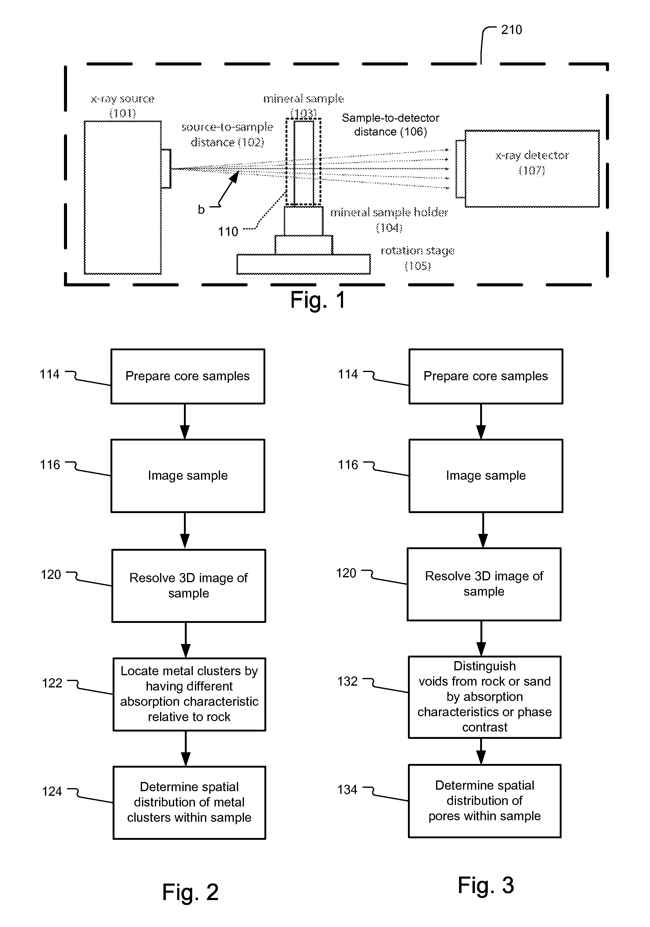
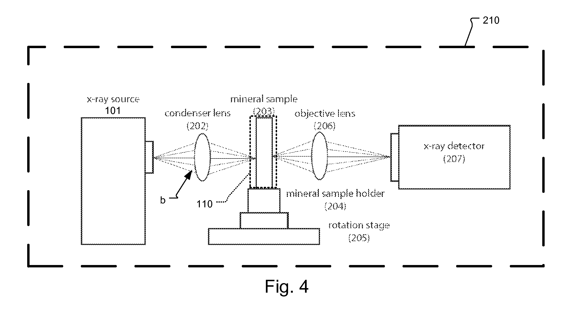
[0017]This disclosure describes a process that uses an x-ray CT system to determine the porosity and / or mineral content of mineral samples. Examples samples include mineral samples such as sandstone, bituminous sand, ore samples, and coal or samples containing precious metals or fluids, such as water or crude oil.

[0018]A basic implementation is shown in FIG. 1. In this configuration, an x-ray source 101 generates x-ray radiation beam b. A mineral sample 103 is placed in the beam path b and the x-ray radiation passing through the sample 103 is recorded by a spatially resolved detector 107, having 1,024×1,024 pixels, for example. The sample is mounted on sample holder 104 with an integrated rotation stage 105 that rotates the sample through a range of −90 degrees and +90 degrees from the optical axis.

[0019]With mineral samples, a high-energy x-ray radiation beam is used with an energy above several keV. This is typically required to penetrate sample with tens of micrometers or greater...


PUM


Abstract

A process to determine the porosity and/or mineral content of mineral samples with an x-ray CT system is described. Based on the direct-projection techniques that use a spatially-resolved x-ray detector to record the x-ray radiation passing through the sample, 1 micrometer or better resolution is achievable. Furthermore, by using an x-ray objective lens to magnify the x-ray image in a microscope configuration, a higher resolution of up to 50 nanometers or more is achieved with state-of-the-art technology. These x-ray CT techniques directly obtain the 3D structure of the sample with no modifications to the sample being necessary. Furthermore, fluid or gas flow experiments can often be conducted during data acquisition so that one may perform live monitoring of the physical process in 3D.

Description

RELATED APPLICATIONS[0001]This application claims the benefit under 35 USC 119(e) of U.S. Provisional Application No. 61 / 043,437, filed on Apr. 9, 2008, which is incorporated herein by reference in its entirety.BACKGROUND OF THE INVENTION[0002]X-ray imaging, including projection and microscopy techniques, have been widely used in medical imaging and industrial inspection since their invention. A particularly important development is the computed tomography (CT) techniques that allow the three dimensional (3D) structure of a sample to be reconstructed from a series of two dimensional (2D) x-ray images acquired at different view angles. Furthermore, recent advances in micro x-ray CT has pushed the 3D resolution to 1 micrometer with direct-projection type imaging systems, while using x-ray objective lenses, such as Fresnel zone plates, 3D images with up to 50 nanometer resolution have been demonstrated.[0003]Mineral samples analysis for mining is important as ores are used in metal pro...

Claims


Application Information

Patent Timeline
IPC IPC(8): G01N23/00
CPCG01N23/046G01N2223/419
Inventor YUN, WENBINGFESER, MICHAELTKACHUK, ANDREICASE, THOMAS A.DUEWER, FREDERICK W.CHANG, HAUYEE
Owner CARL ZEISS X RAY MICROSCOPY
Why Eureka
Try Eureka