
System and method for lesion segmentation in whole body magnetic resonance images

a whole body magnetic resonance and segmentation algorithm technology, applied in image enhancement, image analysis, instruments, etc., can solve the problems of large variation in size, shape, texture, irregular geometry, and manual segmentation is very time-consuming and requires a lot of user interaction, and achieves minimal user interaction and accurate results.

Inactive Publication Date: 2012-04-10
SIEMENS HEALTHCARE GMBH
View PDF4 Cites 29 Cited by
  • Summary
  • Abstract
  • Description
  • Claims
  • Application Information

AI Technical Summary

Benefits of technology

This method enables efficient, robust, and accurate lesion segmentation with minimal user input, providing fast and reliable delineation and visualization of lesions, adaptable to varying image resolutions and anatomical structures, and applicable to multiple imaging modalities.

Problems solved by technology

On the other hand, manual segmentation is very time-consuming and typically requires a lot of user interaction.
For instance, the lesions in the lung and liver may show a smoother geometry and have a lesser intensity variation, however, in general these observations do not hold and lesions typically have an irregular geometry and large variations in size, shape, and texture.

Method used


Image

  • System and method for lesion segmentation in whole body magnetic resonance images
  • System and method for lesion segmentation in whole body magnetic resonance images
  • System and method for lesion segmentation in whole body magnetic resonance images

Examples


Embodiment Construction

[0056]Exemplary embodiments of the invention as described herein generally include systems and methods for an efficient and robust lesion and tumor segmentation algorithm for 3-dimensional whole body magnetic resonance imaging. Accordingly, while the invention is susceptible to various modifications and alternative forms, specific embodiments thereof are shown by way of example in the drawings and will herein be described in detail. It should be understood, however, that there is no intent to limit the invention to the particular forms disclosed, but on the contrary, the invention is to cover all modifications, equivalents, and alternatives falling within the spirit and scope of the invention.

[0057]As used herein, the term “image” refers to multi-dimensional data composed of discrete image elements (e.g., pixels for 2-D images and voxels for 3-D images). The image may be, for example, a medical image of a subject collected by computer tomography, magnetic resonance imaging, ultrasou...


PUM


Abstract

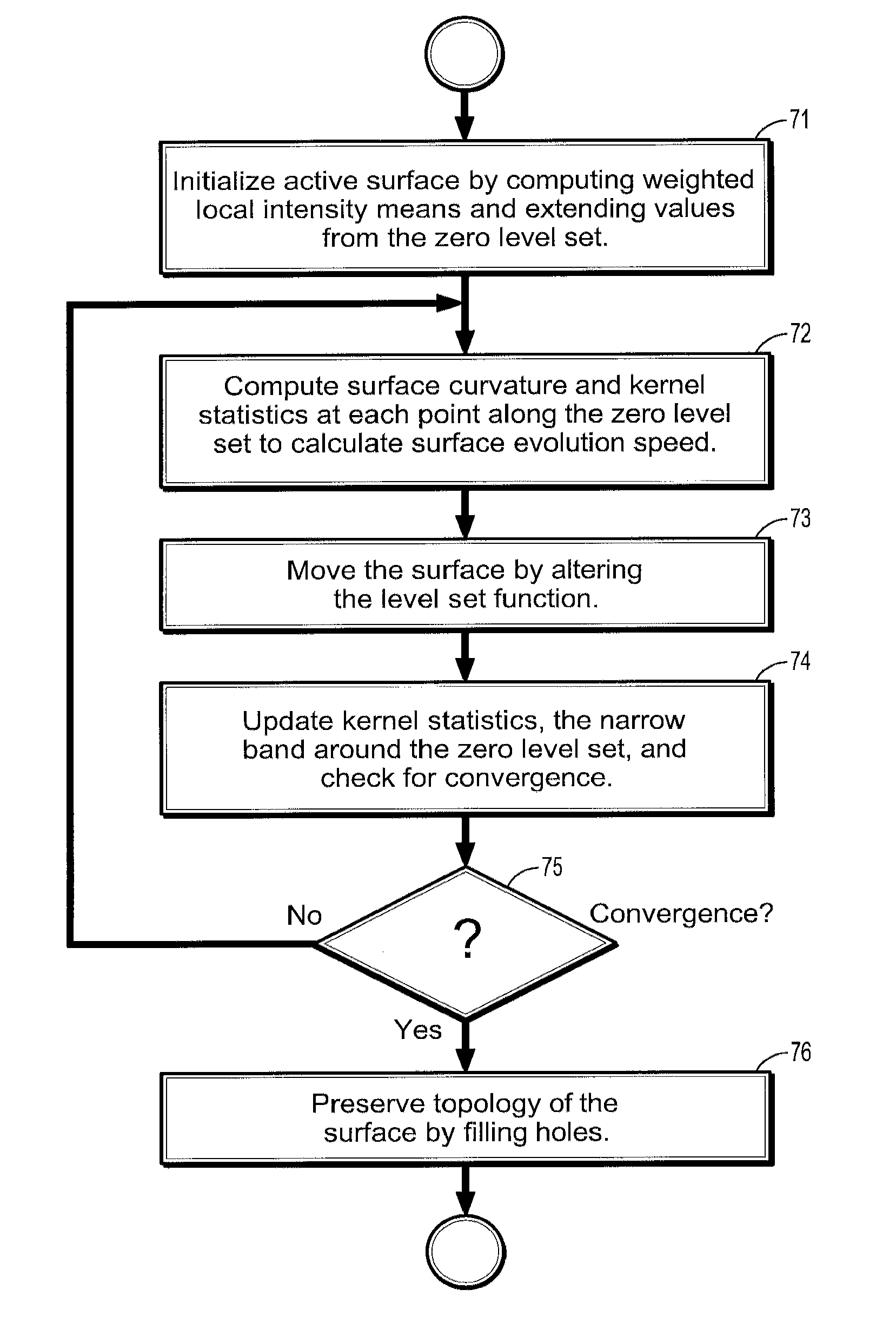
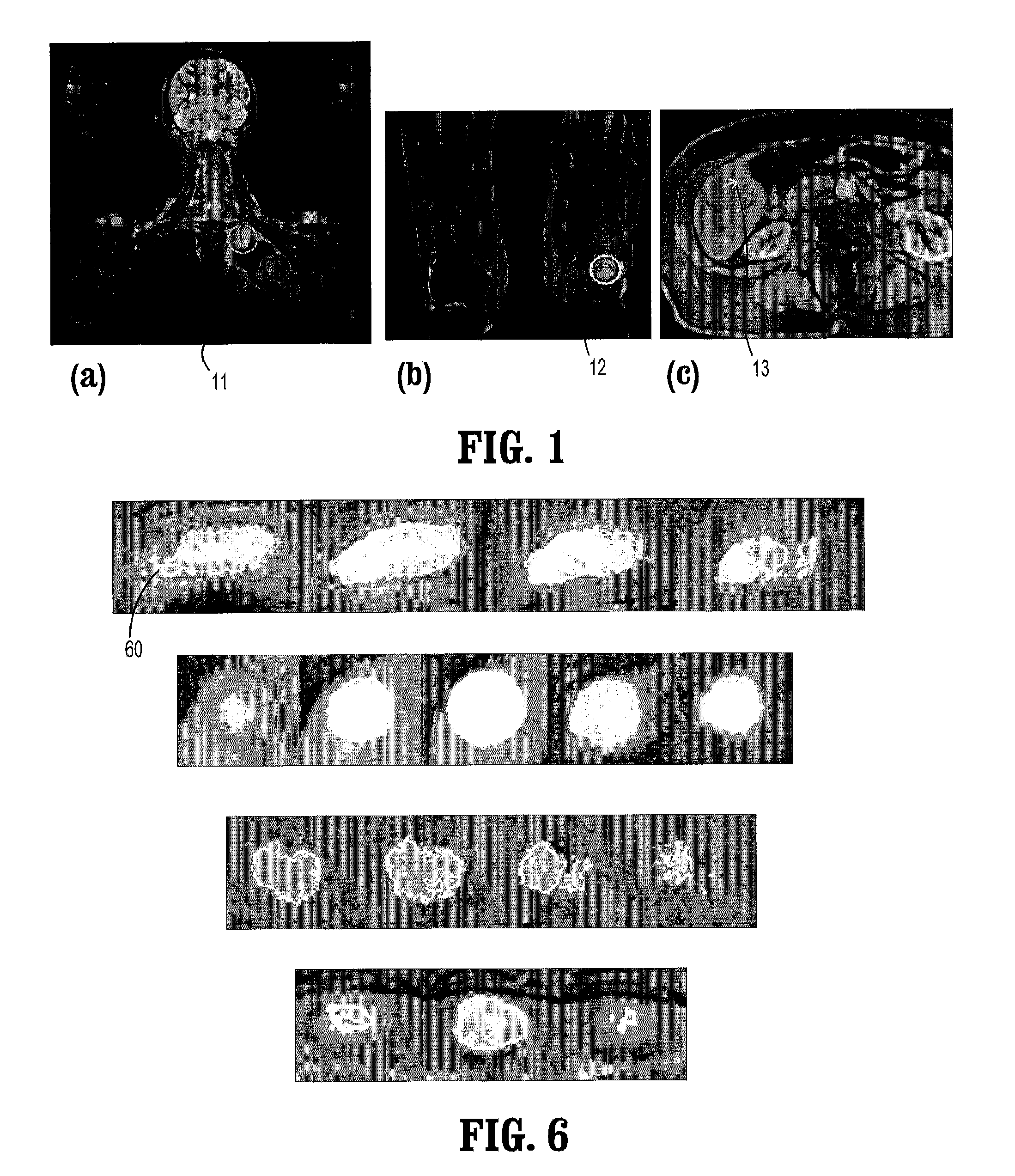
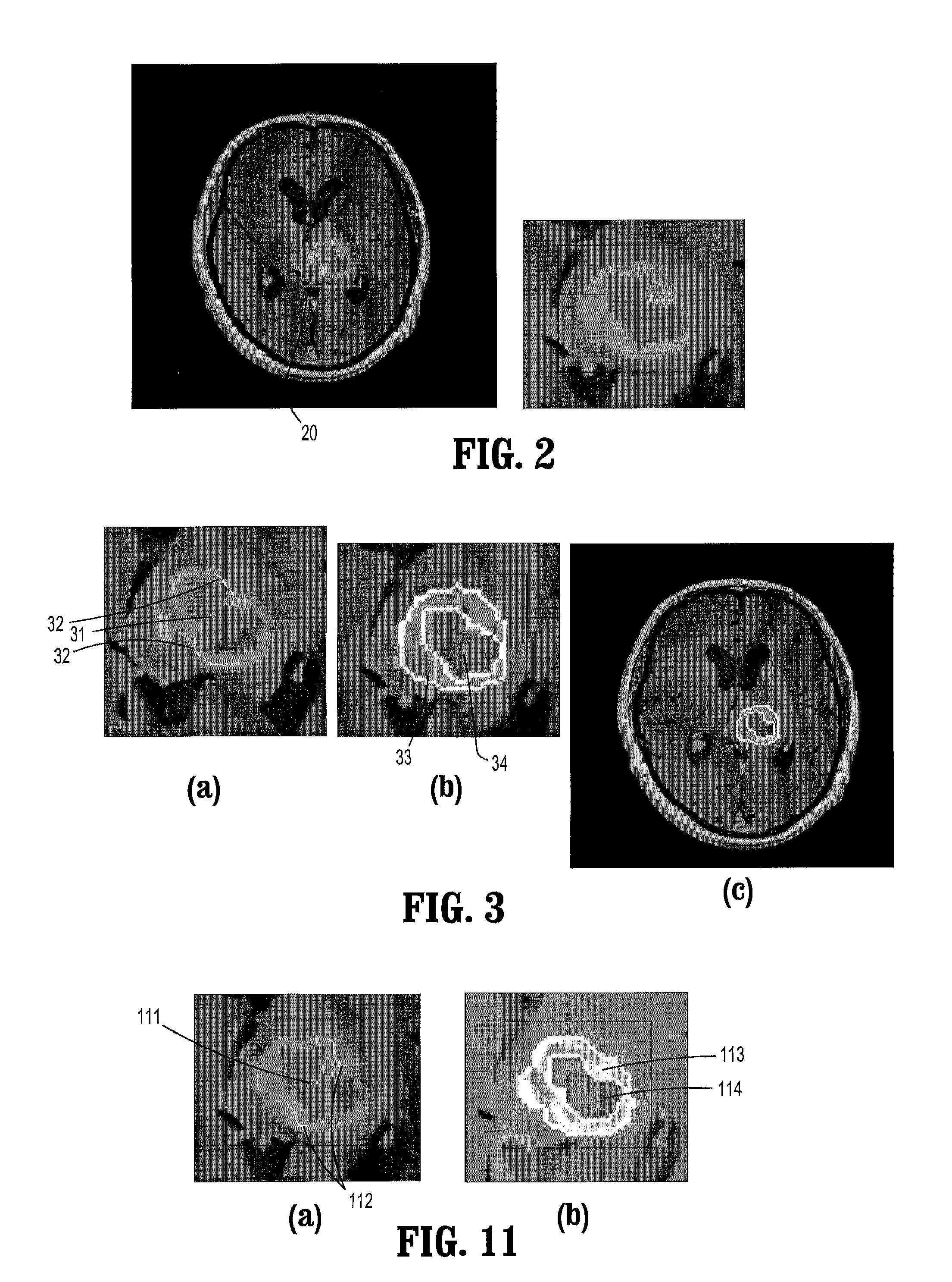
A method for lesion segmentation in 3-dimensional (3D) digital images, includes selecting a 2D region of interest (ROI) from a 3D image, the ROI containing a suspected lesion, extending borders of the ROI to 3D forming a volume of interest (VOI), where voxels on the borders of the VOI are initialized as background voxels and voxels in an interior of the VOI are initialized as foreground voxels, propagating a foreground and background voxel competition where for each voxel in the VOI, having each neighbor voxel in a neighborhood of the voxel attack the voxel, and, if the attack is successful, updating a label and strength of the voxel with that of the successful attacking voxel, and evolving a surface between the foreground and background voxels in 3D until an energy functional associated with the surface converges in value, where the surface segments the suspected lesion from the image.

Description

CROSS REFERENCE TO RELATED U.S. APPLICATIONS[0001]This application claims priority from “A Lesion Segmentation Method for Whole Body Magnetic Resonance Imaging”, U.S. Provisional Application No. 60 / 912,934 of Unal, et al., filed Apr. 20, 2007, “A Multi-Region Lesion Segmentation Method for Magnetic Resonance Imaging”, U.S. Provisional Application No. 60 / 953,708 of Unal, et al., filed Aug. 3, 2007, and “A system for lesion segmentation and analysis in 3D whole body magnetic resonance images”, U.S. Provisional Application No. 60 / 954,588 of Qing, et al., filed Aug. 8, 2007, the contents of all of which are herein incorporated by reference in their entireties.TECHNICAL FIELD[0002]This disclosure is directed to the development of an efficient and robust lesion and tumor segmentation algorithm for 3-dimensional (3D) whole body (WB) magnetic resonance (MR) imaging.DISCUSSION OF THE RELATED ART[0003]The goal of segmentation is to extract the object of interest from background. In medical im...

Claims


Application Information

Patent Timeline
Patent Type & Authority Patents(United States)
IPC IPC(8): G06K9/00G06K9/34
CPCG06K9/342G06K9/6207G06T7/0083G06T7/0089G06K2009/366G06K2209/053G06T2200/04G06T2207/10088G06T2207/20104G06T2207/20144G06T2207/30096G06T7/12G06T7/149G06T7/194G06V10/248G06V10/267G06V10/755G06V2201/032
Inventor UNAL, GOZDESLABAUGH, GREGORY G.FANG, TONGLANKTON, SHAWNCANDA, VALERTHESEN, STEFANQING, SHUPING
Owner SIEMENS HEALTHCARE GMBH
Learn More