Medical image display apparatus, method and program, and recording medium for the program

a technology for medical images and recording media, applied in the field of medical image display apparatus, method and program, and recording media for program, can solve problems such as inability to meet the needs, and achieve the effect of improving image interpretation efficiency and easy image interpretation

Active Publication Date: 2012-04-17
FUJIFILM CORP
View PDF19 Cites 8 Cited by
  • Summary
  • Abstract
  • Description
  • Claims
  • Application Information

AI Technical Summary

Benefits of technology

[0010]The present invention has been made in view of these circumstances, and an object of the present invention is to provide a medical image display apparatus, method and program, and recording medium for the program which can display a plurality of slice images including an abnormal shadow region from a series of slice images constituting a three-dimensional medical image in a mode that is efficient and enables easy image interpretation, thereby contributing improving the efficiency of image interpretation.
[0012]The display mode to provide multi-screen display of the plurality of slice images including the abnormal shadow region is determined based on the result of acquisition of the abnormal shadow region (three-dimensional information for the abnormal shadow region), which eliminates the necessity for users (doctors) to perform an operation to set a display mode, and enables display of the plurality of slice images including the abnormal shadow region efficiently in a display mode enabling easy image interpretation.
[0028]According to the present invention, it is possible to automatically display a plurality of slice images including an abnormal shadow region from a series of slice images constituting a three-dimensional medical image in a display mode that is efficient and enables easy image interpretation, thereby enabling contributing to the improvement of the efficiency of image interpretation.

Problems solved by technology

However, the medical image management and display system described in Patent Document 1 has a problem in that it requires a user to provide display mode information to each medical image in advance, which is cumbersome.
Furthermore, in recent years, since numerous slice images are taken at one time by means of a multi-slice X-ray CT apparatus or the like, efficient image interpretation is needed, but the inventions described in Japanese Patent Application Laid-Open No. 2005-334634 and Patent Application Laid-Open No. 2002-143095 cannot meet that need.

Method used


Image

  • Medical image display apparatus, method and program, and recording medium for the program
  • Medical image display apparatus, method and program, and recording medium for the program
  • Medical image display apparatus, method and program, and recording medium for the program

Examples


Embodiment Construction

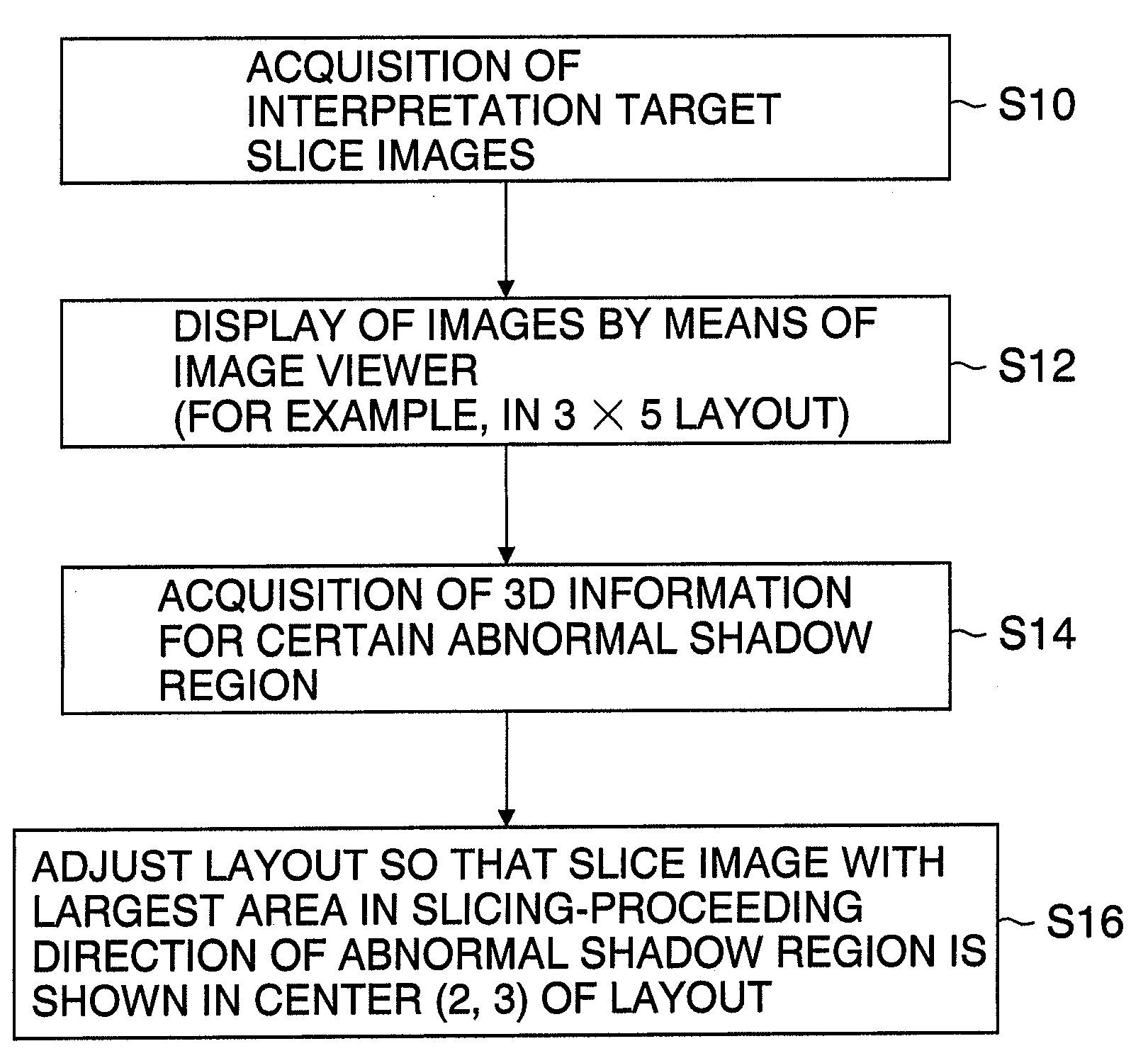
[0043]Hereinafter, preferred embodiments of a medical image display apparatus, method and program, and recording medium for the program according to the present invention will be described with reference to the attached drawings.

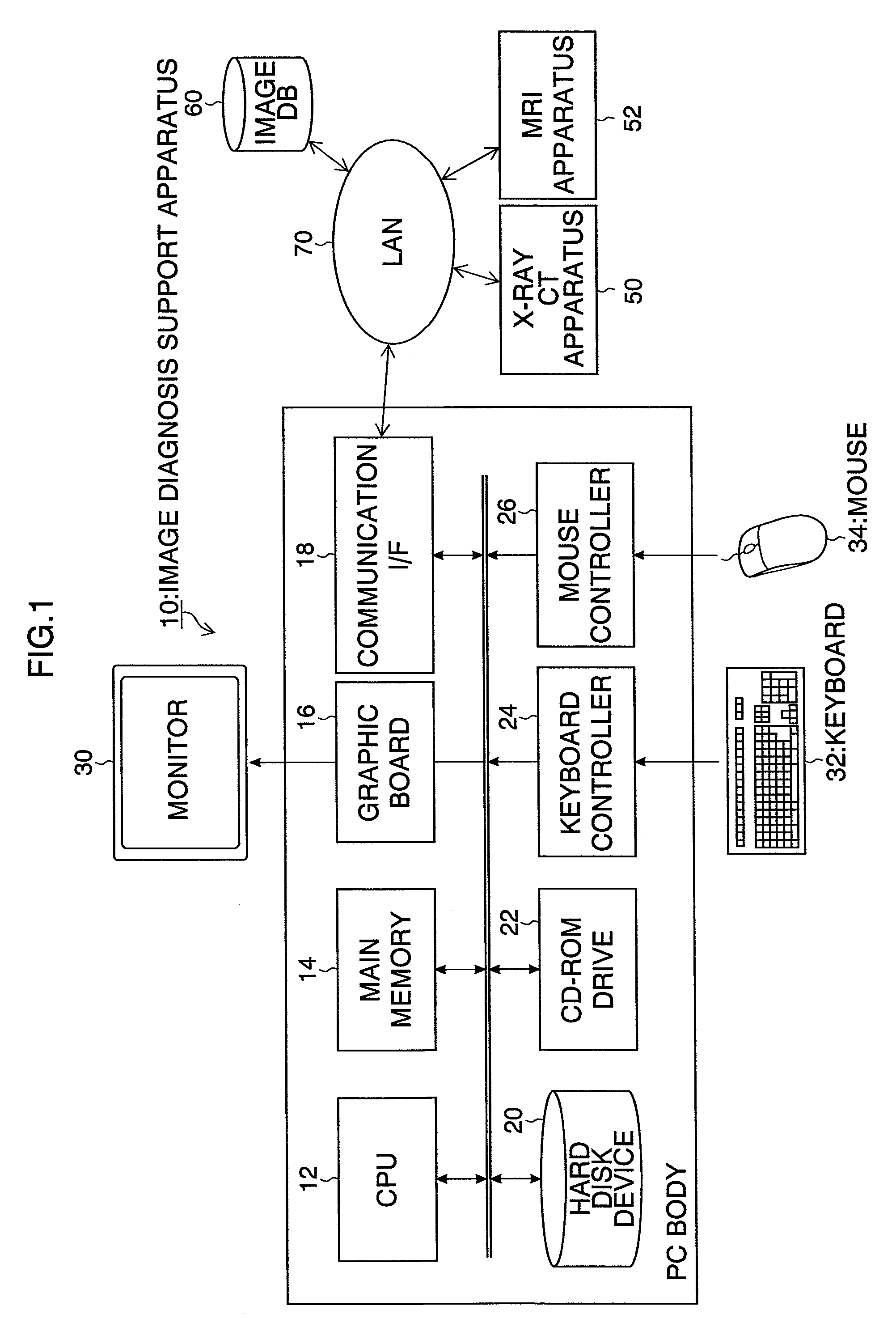
[0044]FIG. 1 is a system configuration diagram of a medical image management system, such as a PACS, that includes a medical image display apparatus according to the present invention.

[0045]This medical image management system mainly includes a medical image display apparatus 10, which is operated by diagnostic radiologists, clinicians, etc., an X-ray CT apparatus 50, an MRI apparatus 52, image database (image DB) 60, and a network 70, such as an in-house LAN, that connects these devices.

[0046]The medical image display apparatus 10, which is constituted by a personal computer (PC), mainly includes a central processing unit (CPU) 12 that controls the operation of the respective components, main memory 14 that stores a control program for the apparatus and bec...


PUM


Abstract

According to the medical image display apparatus of an aspect of the present invention, the display mode to provide multi-screen display of the plurality of slice images including the abnormal shadow region is determined based on the result of acquisition of the abnormal shadow region (three-dimensional information for the abnormal shadow region), which eliminates the necessity for users (doctors) to perform an operation to set a display mode, and enables display of the plurality of slice images including the abnormal shadow region efficiently in a display mode enabling easy image interpretation.

Description

BACKGROUND OF THE INVENTION[0001]1. Field of the Invention[0002]The present invention relates to a medical image display apparatus, method and program, and recording medium for the program. More specifically, the present invention relates to a technique for displaying on a multi-screen a series of slice images taken by means of an X-ray CT apparatus or an MRI apparatus, etc.[0003]2. Description of the Related Art[0004]Conventionally, a medical image management and display system configured so that an image file containing medical images is provided with display mode information having information for a split screen and medical image frame positions, and when displaying the medical images contained in this image file on a viewer, the screen of the viewer is split according to the split screen information included in the display mode information, and the medical images are displayed at the corresponding positions in the split screen according to the frame position information included...

Claims


Application Information

Patent Timeline
Patent Type & Authority Patents(United States)
IPC IPC(8): G06K9/00
CPCA61B6/032A61B6/463A61B6/466A61B6/566G06F19/321G06F19/3406G06T7/0012A61B5/055G06T2200/24G06T2207/10072G06T2207/30096G16H30/20G16H30/40G16H40/63G16H50/20
Inventor LI, YUANZHONG
Owner FUJIFILM CORP
Why Eureka
Try Eureka