System and method for intracorporeal elastography

a technology of intracorporeal elastography and system and method, which is applied in the direction of using reradiation, vibration diagnostics, sensors, etc., can solve the problems of limited technique, high attenuation of shear waves in the body, and limited range of palpation to superficial organs and the sensitivity of tactile perception

Active Publication Date: 2012-07-17
SUNNYBROOK HEALTH SCI CENT
View PDF13 Cites 4 Cited by
  • Summary
  • Abstract
  • Description
  • Claims
  • Application Information

AI Technical Summary

Benefits of technology

[0011]In accordance with one aspect of the invention, an apparatus for intracorporeal elastography includes an intra-luminal vibratory member configured to be positioned within a lumen of an imaging subject and configured to impart mechanical energy into tissue of the lumen. An acoustic energy source is also included and externally coupled to the vibratory member, thereby causing the vibratory member to generate shear waves within the subject.

Problems solved by technology

Unfortunately, palpation is mostly limited to superficial organs and to the sensitivity of tactile perception.
The technique is limited to measuring static elastic properties and is dependent on the operator.
Unfortunately, shear waves are highly attenuated in the body and are typically limited to less than 10 cm of penetration at frequencies between 50-60 Hz.
In addition, the low frequencies used to achieve sufficient penetration result in longer shear wavelengths in tissue that result in lower spatial resolution elastograms.
While this approach provides an initial step in the development of intracorporeal MRE, it suffers from the disadvantage of requiring that the ultrasonic transducer be mounted and powered at the distal end of the catheter (i.e. the end of the catheter that resides within the subject during imaging).
This requirement adds significant complexity to the design of the system and necessitates powering the transducer by electrical leads integrated axially along the catheter.
Such an arrangement is costly to produce, may be prone to an increased failure rate, and may also generate unwanted signal artifacts.

Method used


Image

  • System and method for intracorporeal elastography
  • System and method for intracorporeal elastography
  • System and method for intracorporeal elastography

Examples


Embodiment Construction

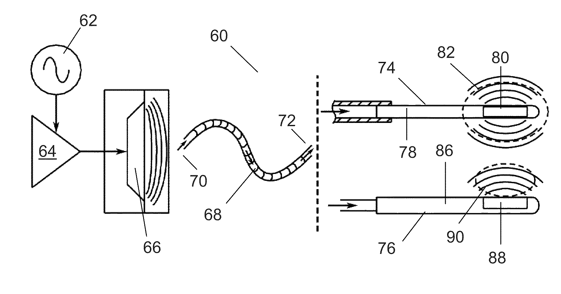
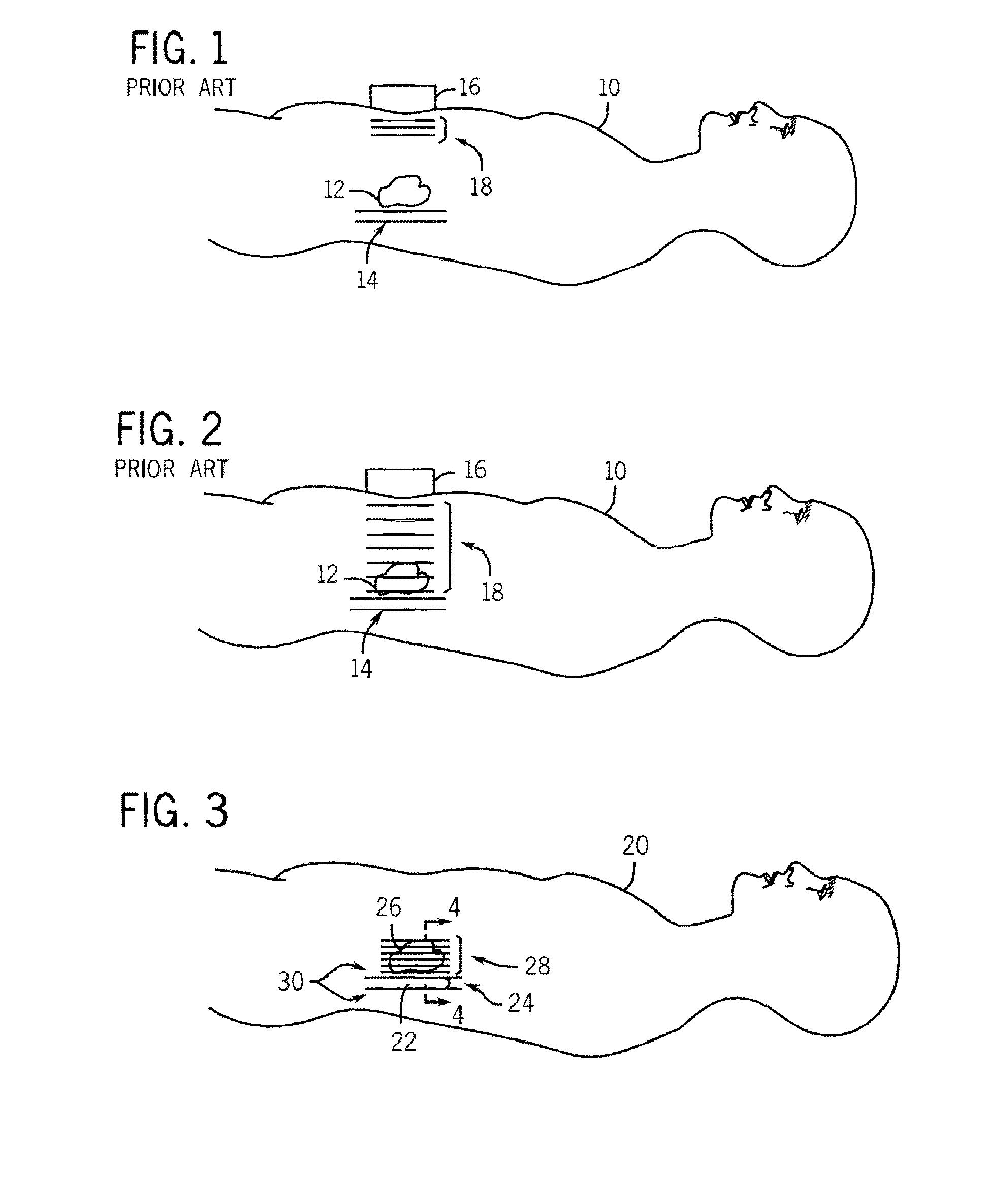
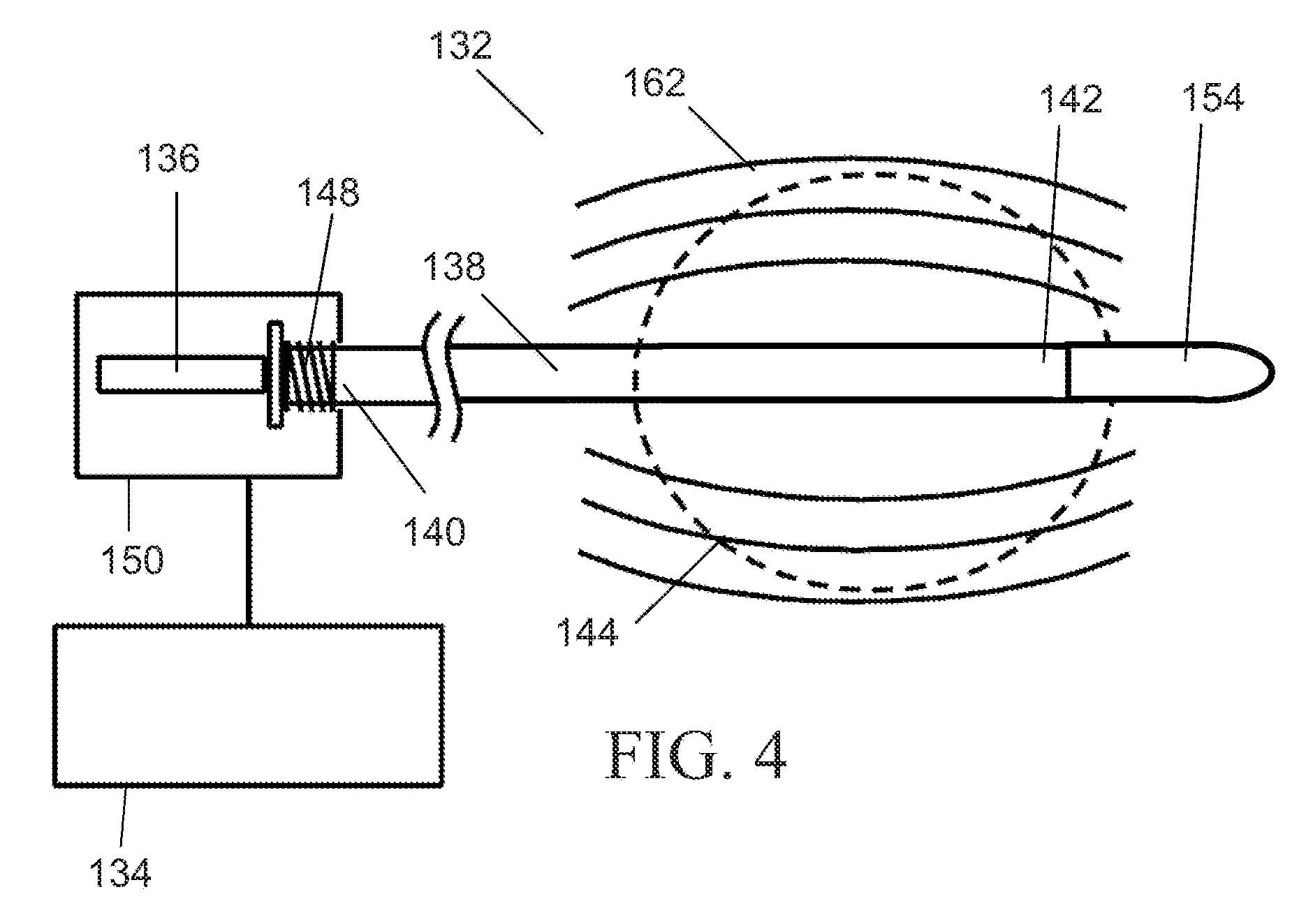
[0045]Generally speaking, the systems described herein are directed to an intra-luminal vibratory device for magnetic resonance elastography. As required, embodiments of the present invention are disclosed herein. However, the disclosed embodiments are merely exemplary, and it should be understood that the invention may be embodied in many various and alternative forms. The Figures are not to scale and some features may be exaggerated or minimized to show details of particular elements while related elements may have been eliminated to prevent obscuring novel aspects. Therefore, specific structural and functional details disclosed herein are not to be interpreted as limiting but merely as a basis for the claims and as a representative basis for teaching one skilled in the art to variously employ the present invention. For purposes of teaching and not limitation, the illustrated embodiments are directed to an intra-luminal vibratory device for magnetic resonance elastography.

[0046]As...


PUM


Abstract

A system and method for intracorporeal elastography include an intra-luminal vibratory member configured to be positioned within a lumen of an imaging subject and configured to impart mechanical energy into tissue of the lumen. In a preferred embodiment, an external piezoelectric energy source is included and coupled to the vibratory member and configured to cause the vibratory member to longitudinally vibrate, thereby generating shear waves for use with magnetic resonance elastography.

Description

CROSS-REFERENCE TO RELATED APPLICATIONS[0001]This application claims the benefit of U.S. Provisional Application No. 61 / 118,250, filed Nov. 26, 2008, and U.S. Provisional Application No. 61 / 090,317, filed Aug. 20, 2008, which are each herein incorporated in their entirety.FIELD OF THE INVENTION[0002]The present invention relates generally to elasticity imaging for medical diagnosis and treatment evaluation and, more particularly, to the measurement of the stiffness distribution in a region of tissue using an intra-luminal device placed within a body cavity or organ to produce mechanical vibration of adjacent tissue.BACKGROUND OF THE INVENTION[0003]Many disease processes such as solid tumors and liver fibrosis manifest themselves as changes in tissue mechanical properties. Manual palpation of tissues is an important clinical tool for the detection of disease within the body. Unfortunately, palpation is mostly limited to superficial organs and to the sensitivity of tactile perception....

Claims


Application Information

Patent Timeline
Patent Type & Authority Patents(United States)
IPC IPC(8): G01V3/00
CPCG01R33/28A61B8/485G01R33/56358G01R33/563A61B5/0051A61B5/055
Inventor CHOPRA, RAJIVBRONSKILL, MICHAEL JOHNFORGHANIAN-ARANI, ARVIN
Owner SUNNYBROOK HEALTH SCI CENT
Why Eureka
Try Eureka