
Flaccid tubular membrane and insertion appliance for surgical intubation

a tubular membrane and intubation technology, applied in the field of medical science, can solve the problems of urethral obstruction, substantial burden on the delivery of effective care through the healthcare system, and insufficient flexibility of protective sheath,

Active Publication Date: 2013-01-01
HARMON SR JAMES V
View PDF38 Cites 0 Cited by
  • Summary
  • Abstract
  • Description
  • Claims
  • Application Information

AI Technical Summary

Benefits of technology

The sheath facilitates easier and faster catheter placement, reduces tissue trauma, and decreases healthcare costs by enabling non-urologists to navigate obstructions efficiently without cystoscopes, while maintaining access for fluid drainage and instrument insertion.

Problems solved by technology

While most placements proceed without problems, typically about ten percent of urinary catheter placements are difficult, causing a substantial burden on the delivery of effective care through the healthcare system.
The most common problem is tetany, a spasm of the external urinary sphincter or stricture of the urethra.
Stones, and even clots descending from the bladder, also constitute urethral obstructions.
The cost to the healthcare system, hospitals, clinics and doctors' offices is substantial.
In addition, the delay in servicing urological catheter patients in a timely manner constitutes poor medical efficiency, delivery and control.
When difficulty is encountered, the resulting frustration among healthcare professionals, especially nurses, physician extenders and physician assistants, creates a very real feeling of ineffectiveness on the part of these healthcare workers, to say nothing of the dissatisfaction on the part of the patients caused by the delay and added discomfort.
While the dollar cost to the healthcare system is not the only concern, such elements as added labor and material costs, time delays for patient rectification, excess space and equipment required, catheter kit value, nurse technician and physician costs constitute an expense to the healthcare system of surprising proportions.
Of these, about 15,000 are difficult.
Moreover, the additional space and equipment amounts to at least $800 million per year for a total added cost of about $1.5 billion per year.
The repeated insertion and removal of instruments often requires a significant amount of force.
This can of course traumatize the tissue.
In addition, the manipulation of a surgical instrument or other object that is partially or completely inserted into the body can also result in damage to the surrounding tissue.
The insertion and removal of each of these other instruments increases the chances for traumatizing or injuring surrounding tissue or even creating a false passage and losing access.
In short, tissue trauma can result from retrograde or antegrade passage instrumentation or removal of foreign bodies. many endoscopic, percutaneous or laparoscopic instruments have a relatively small diameter working channel which limits the size of biopsy specimens.
The small size limits the removal of such specimens or foreign bodies by necessitating multiple insertions and withdrawals.
This prolongs the operation and is an additional source of tissue trauma.
Catheters, now in common use, however have thick walls, typically about 2 mm or more in thickness, which have limited capacity to expand so that some instruments can be passed through the lumen of the catheter only with great difficulty or not at all.
However there is no way to hold the sheath in place one inserted.
However the insertion and positioning of a thin sheath has been difficult to accommodate largely because of the thin flexible, flaccid walls.

Method used


Image

  • Flaccid tubular membrane and insertion appliance for surgical intubation
  • Flaccid tubular membrane and insertion appliance for surgical intubation
  • Flaccid tubular membrane and insertion appliance for surgical intubation

Examples


Embodiment Construction

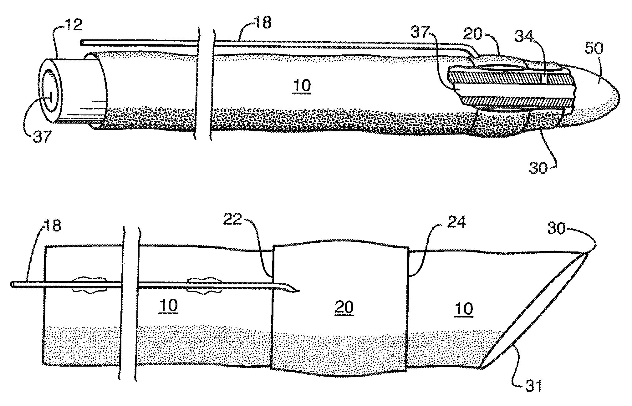
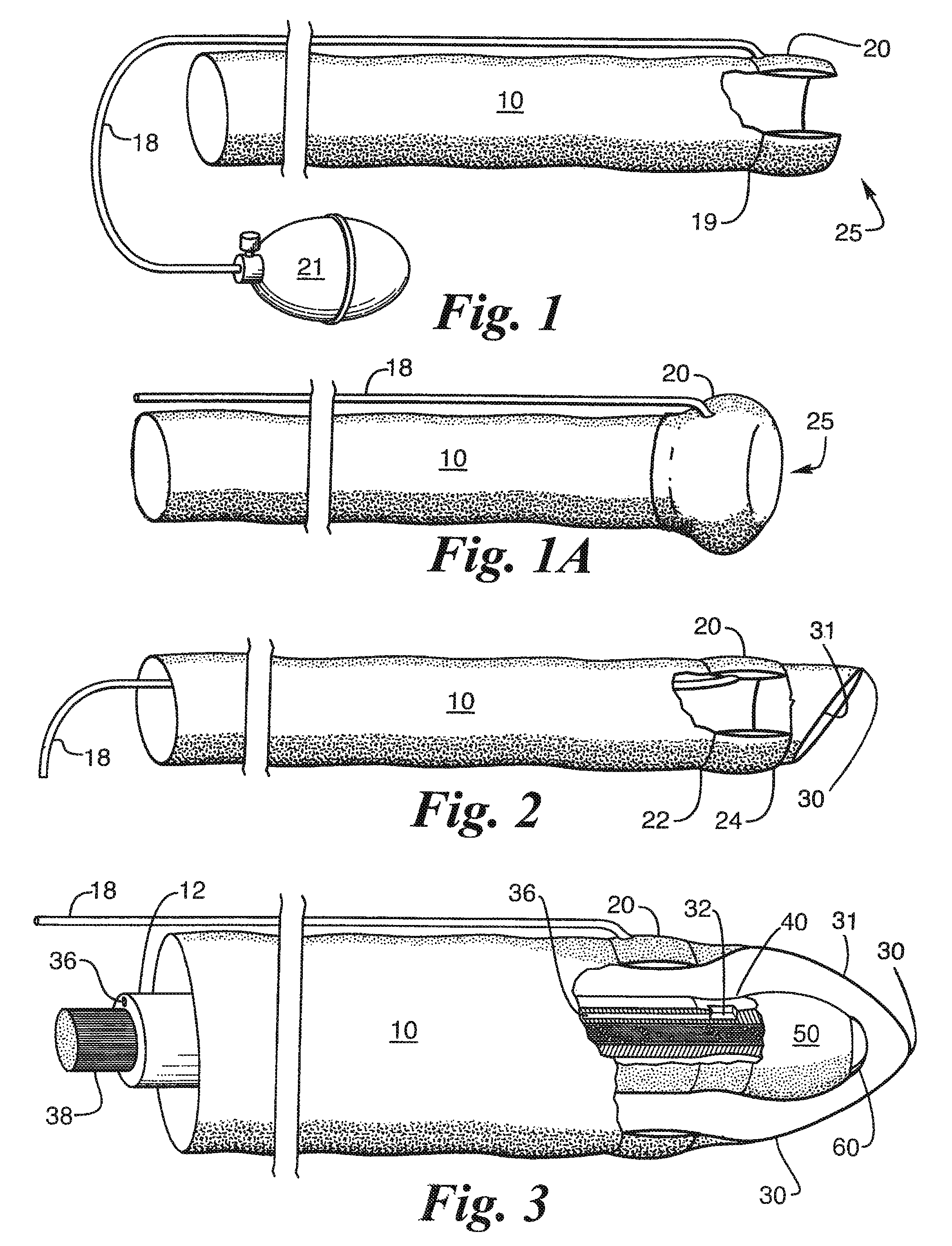
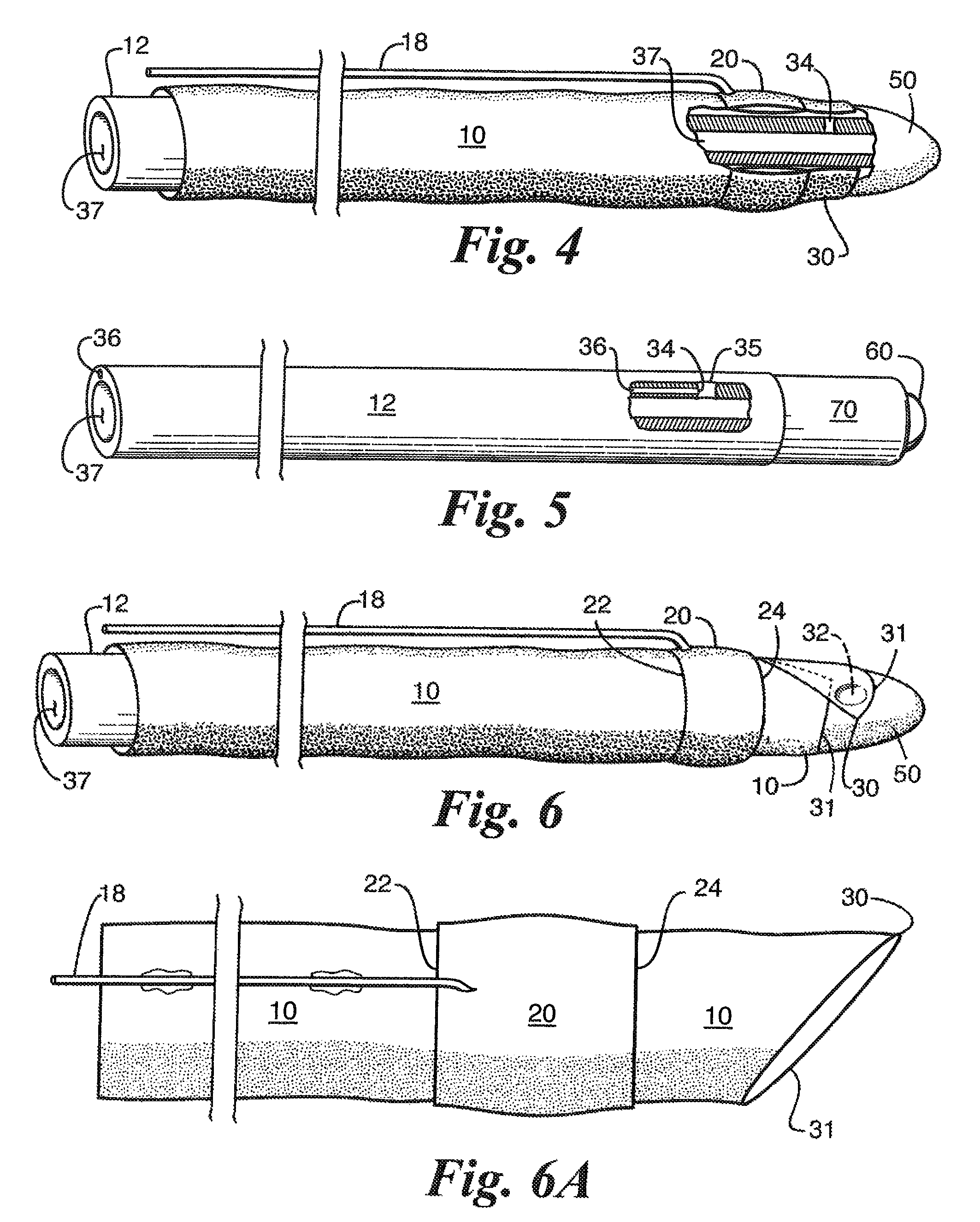
[0036]A sheath 10 in accordance with the present invention is a flexible flaccid and membranous tube with an annular balloon 20 at one end. The sheath functions to protect tissue in a body orifice during subcutaneous surgical procedures or intubations. A very thin sheath is preferred to be more easily inserted and to accept a wider range of surgical tool diameters by being able to expand and contract to the instrument size of the tools being used by the surgeon during the procedures and to be more easily removed after a procedure since it is of a smaller cross section when it has thinner membranous walls.

[0037]As shown in FIG. 1 and FIG. 1A a sheath 10 has a balloon 20 formed at the distal end 25 of the sheath. The balloon may be formed by simply manually expanding the free distal end then everting and sliding the sheath material back toward the opposite end and adhesively bonding or otherwise attaching the free edge of the sheath to the underlying material of the sheath at 19 near ...


PUM


Abstract

A thin sheath of flexible, flaccid material is introduced through openings in the body of an animal or human patient to permit surgical tools or fluids to be inserted and removed, or debris removed, during surgeries anywhere in the body to protect the tissue of the body from the instruments used. The sheath is inserted by an obturator which holds the sheath thereon by suction through an aperture in the obturator at its distal end. Once the sheath is inserted, suction is removed to release the sheath. Then a balloon on the distal end of the sheath is expanded to hold the sheath in place in the body while the obturator is removed. The sheath then remains in place with the balloon holding the distal end in the open position to enable fluids to flow through the sheath. After a procedure, the balloon is deflated and the sheath removed.

Description

CROSS REFERENCE TO RELATED APPLICATION[0001]This application is a divisional of application Ser. No. 11 / 853,666, filed Sep. 11, 2007 now U.S. Pat. No. 7,862,542, and entitled “Flaccid Tubular Membrane and Insertion Appliance for Surgical Intubation”.[0002]Applicant claims the benefit of Provisional Application Ser. No. 60 / 834,484 filed Sep. 11, 2006 having the same title which is incorporated herein by reference.BACKGROUND OF THE INVENTION[0003]1. Field of the Invention[0004]This invention relates the medical arts and more particularly to a flexible protective sheath for subcutaneous insertion into a body such as for facilitating urological, and gastrological procedures.[0005]2. Description of the Related Art[0006]The invention can be used in a variety of surgical fields such as urology and gastroenterology. The invention will be described by way of example in connection with the field of urology. One of the most common urological procedures, both historically and in current practic...

Claims


Application Information

Patent Timeline
Patent Type & Authority Patents(United States)
IPC IPC(8): A61B1/04A61B1/00
CPCA61B1/00135A61B1/00154A61B17/3421A61M25/0017A61M25/01A61M25/0662A61M25/10A61B17/3431A61B2017/3456A61B2017/3486A61B2019/5217A61M2025/0233A61M2025/0293A61B2090/3614
Inventor HARMON, SR., JAMES V.
Owner HARMON SR JAMES V
Learn More