Supercharge Your Innovation With Domain-Expert AI Agents!

Apparatus and method for processing image information captured over time at plurality of positions in subject body

a technology of image information and apparatus, applied in the field of apparatus and method for processing image information captured over time at plurality of positions in the subject body, can solve the problems of not easy to locate image information, not easy to comprehend such a large number, and potentially enormous number of image information acquired by imaging during the period

Active Publication Date: 2013-02-05
OLYMPUS CORP
View PDF13 Cites 2 Cited by
  • Summary
  • Abstract
  • Description
  • Claims
  • Application Information

AI Technical Summary

Benefits of technology

Facilitates the easy comprehension and location of specific image information by visually correlating image data with positional information, enhancing diagnostic efficiency and reducing the time required to analyze large datasets.

Problems solved by technology

However, in such a capsule endoscope, unlike conventional endoscopes, because the observation period or examination period is the time period from being swallowed by the patient until being naturally passed out of the body, the observational period and examination period may last for a long time, more than 10 hours for example, and therefore the number of image information acquired by imaging during the period is potentially enormous.
In these circumstances, during the stages of a diagnosis etc., it is not easy to comprehend such a large number of image information in a short period, and it is also not an easy task to locate the image information of a part intended to be focused on, or more specifically image information exclusively of an organ intended to be diagnosed or image information related to an image capturing an ailing part etc. from an enormous number of image information.
Therefore, by the device and method described in the document alone, it is in practice difficult to realize display of the positions of the image and the capsule on a single monitor.
However, although such a display by a progress bar can indicate time progress or the percentage of processing completed, the location currently being processed of the processed subject cannot be indicated.
Likewise, the technique disclosed in the above Patent Document 1, does not provide any system to indicate the processing status that is the location in the luminal organs of the body, when performing image processing such as extracting a particular image from a number of images.

Method used


Image

  • Apparatus and method for processing image information captured over time at plurality of positions in subject body
  • Apparatus and method for processing image information captured over time at plurality of positions in subject body
  • Apparatus and method for processing image information captured over time at plurality of positions in subject body

Examples


first embodiment

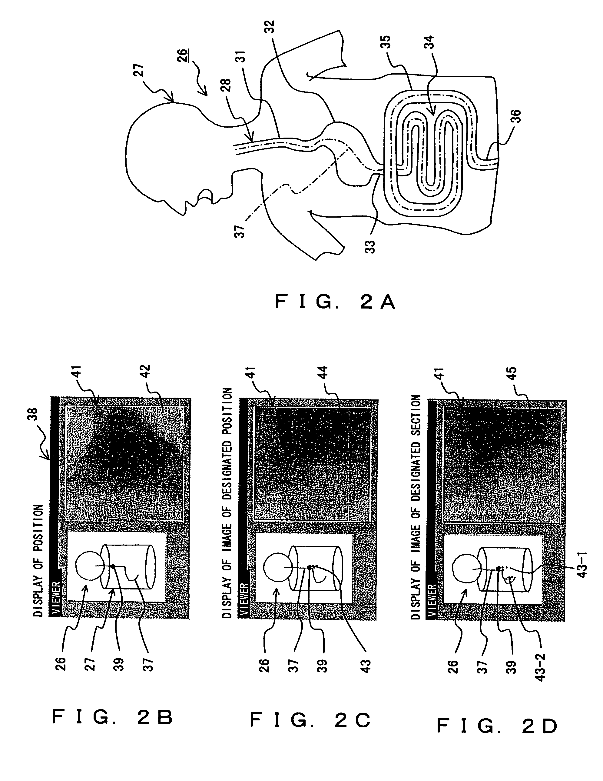
[0084]FIG. 2A, FIG. 2B, FIG. 2C and FIG. 2D are diagrams of the subject internal model 26 of the subject individual 4 and examples of an image 42, both of which are displayed on the same display screen 38 of the monitor device 21 of the workstation in the capsule endoscope image filing system relating to the present invention in terms of the FIG. 2A is a diagram showing the subject internal model of the subject individual 4 displayed on a display screen of the monitor device 21 and FIG. 2B, FIG. 2C and FIG. 2D are diagrams showing examples of images displayed on the same display screen of the monitor device 21 as the subject internal model. FIG. 2B, FIG. 2C and FIG. 2D simplify the subject internal model shown in FIG. 2A for the convenience of the explanation.

[0085]As shown in FIG. 2A, both the subject body model 27 of the subject individual 4 and a digestive organ model 28 are indicated as a frame format of a two-dimensional diagram as a subject internal model 26 of the subject in...

second embodiment

[0122]Following the above process, the processing operation of the image processing in the capsule endoscope image filing system in the second embodiment is explained.

[0123]FIG. 5 is a flowchart explaining the operation of the image processing in the second embodiment. This image processing is also processing performed by the CPU of the main device 19 based on the instruction input by a doctor or a nurse from the keyboard 22 or the mouse 23 of the workstation 7 shown in FIG. 1. In this case also, the operation explained in the first embodiment is performed in advance of this processing.

[0124]In FIG. 5, processing S1-S3, S4, and S5 are the same as the processing S1-S3, S4 and S5, respectively, of the first embodiment explained with the flowchart of FIG. 3. In the present embodiment, processing S31 is performed between the processing S3 and the processing S4.

[0125]Specifically, after the processing of displaying the focus position information (S3), a doctor or a nurse designates extra...

third embodiment

[0148]FIG. 7 is a flowchart explaining another example of the operation of the image processing in the In this processing, processing S32 and processing S33 are performed instead of the processing S6 shown in FIG. 6.

[0149]In other words, following the display of the first focus position information (S4) and the display of the image corresponding to the focus position information (S5), it is determined (S32) whether or not the first position of the focus position information is designated.

[0150]This processing is processing, in response to the input operation of the “section designation” menu or the “section designation” button by a doctor or a nurse operating the mouse 23, by the subsequent operation of the mouse 23, to determining whether or not any of the positions on the display of the trajectory path 37 of the subject internal model 26 is designated as the position to be focused on first in the above designated section, that is the first position.

[0151]When the first position, ...


PUM


Abstract

An image captured by successive imaging over time by using a capsule endoscope is correlated with the imaging position and compiled into a database. The left side of a display screen of a monitor device displays a subject internal model, a trajectory path of the capsule endoscope, and a position mark indicating the imaging position. An image corresponding to the position of the position mark is displayed in an image display area on the right side. When a position mark on the trajectory path is designated by a mouse, the corresponding image is displayed. It is also possible to designate a section for which the image corresponding to the position mark is displayed or to designate a position mark where an unusual image is captured.

Description

CROSS REFERENCE TO RELATED APPLICATIONS[0001]This application is a continuation application of international PCT application No. PCT / JP2004 / 018170 filed on Dec. 6, 2004.[0002]This application claims benefit of Japanese Applications No. 2003-408233, filed Dec. 5, 2003, and NO. 2004-28579, filed Feb. 4, 2004, the contents of which are incorporated by this reference.BACKGROUND OF THE INVENTION[0003]1. Field of the Invention[0004]The present invention relates to a technique such as capsule endoscopy, which moves autonomously or heteronomously within the body of a subject and performs display processing of a plurality of information imaged over time, and to a maintenance technique of successively shot image data captured by such an automatic traveling type imaging device.[0005]2. Description of the Related Art[0006]Lesions of luminal organs such as the esophagus and gastrointestinal organs include bleeding, tissue damage, vascular anomalies, Crohn's disease, polyps and so forth, for exam...

Claims


Application Information

Patent Timeline
Patent Type & Authority Patents(United States)
IPC IPC(8): A62B1/04H04N7/18A61B1/273A61B1/31A61B5/06A61B5/07
CPCA61B1/00009A61B1/0005A61B1/041A61B1/273A61B5/06A61B1/00016A61B1/31A61B5/061
Inventor HIRAKAWA, KATSUMIHARA, MASANAO
Owner OLYMPUS CORP
Features
  • R&D
  • Intellectual Property
  • Life Sciences
  • Materials
  • Tech Scout
  • Unparalleled Data Quality
  • Higher Quality Content
  • 60% Fewer Hallucinations
Learn More